
FIGURE
Fig. 4
- ID
- ZDB-FIG-090511-18
- Publication
- Willett et al., 1997 - Expression of zebrafish rag genes during early development identifies the thymus
- Other Figures
- All Figure Page
- Back to All Figure Page
Fig. 4
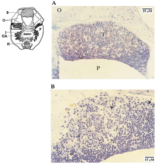
The thymus at 1 month. (A) The thymic epithilium is continuous with the pharyngeal epithilium (location of section is shown in diagram at left). (B) At higher magnification, thymocytes appear as groups of packed cells between nonlymphoid cells, including epithelial cells. Photos are of plastic sections stained with toluidine blue. B, brain; GA, gill arches; H, heart; I, intestine; O, otic vesicle; P, pharynx; T, thymus. Lymphoblasts, solid arrows marked ‘‘1’’; small lymphocytes, open arrows marked ‘‘2’’; epithelial cells, double arrowheads marked ‘‘3.’’ |
Expression Data
Expression Detail
Antibody Labeling
Phenotype Data
Phenotype Detail
Acknowledgments
This image is the copyrighted work of the attributed author or publisher, and
ZFIN has permission only to display this image to its users.
Additional permissions should be obtained from the applicable author or publisher of the image.
Reprinted from Developmental Biology, 182(2), Willett, C.E., Zapata, A.G., Hopkins, N.A., and Steiner, L.A., Expression of zebrafish rag genes during early development identifies the thymus, 331-341, Copyright (1997) with permission from Elsevier. Full text @ Dev. Biol.