|
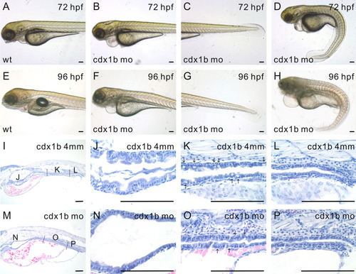
cdx1b antisense morpholino oligonucleotide (MO) knockdown analyses. A-D: Wild type (A) and cdx1b MO-injected (B-D) 72 hr post-fertilization (hpf) embryos. E-H: Wild-type (E) and cdx1b MO-injected (F-H) 96-hpf embryos. I-L: Histological analyses of paraffin sagittal sections of a 96-hpf cdx1b-4mm-MO-injected embryo. Higher magnifications depicting the intestinal bulb (J), mid-intestine (K), and posterior intestine (L). M-P: Histological analyses of paraffin sagittal sections of a 96-hpf cdx1b morphant. Higher magnifications depicting the intestinal bulb (N), mid-intestine (O), and posterior intestine (P). Arrows indicate locations of goblet cells in the mid-intestine (K,O). Scale bars = 100 μm.
|