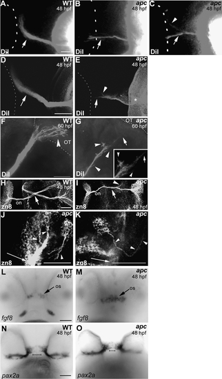
Axon pathfinding defects in apc mutants. (A–E) Whole-eye fills with DiI at 48 hpf show that in most apc mutants, the ON does not cross the OC properly. In 25% of embryos, the ON does not cross at the OC and displays branching axons (arrow in B). In 50% of mutants, a portion of the ON projects toward the optic tract (arrowhead in C), while the remaining axons project into the contralateral ON (arrow in C). In some of the apc mutants with misprojecting ON into the contralateral ON, the ON from the opposite eye appears to initiate correct projection to the OT (arrowhead in E). In addition, axons regularly branch off after exiting the eye (asterisk in E). Ventral view. The outline of the contralateral eye is indicated with dashed lines. (F, G) Lateral view of embryos with DiI-labeled ON in wild-type (F) and apc mutant (G) at 60 hpf. Overlay of fluorescence and transmission images. Anterior to the left. In wild-type embryos (F), the ON terminates in the OT (arrowhead). In apc mutants (G), some RGC axons branch off the ON (arrowheads) and the ON does not reach the tectum (arrow). Inset ON magnification. Dashed lines indicate boundary between ventral midbrain and OT. (H–K) Dorsal flatmount view of RGCs and their projections labeled with α-zn8 antibody at 48 hpf shows pathfinding defects at the OC (arrow), whereas some axons continue along the optic tract (arrowhead). Box in (I) shows magnified area in (J, K). (J, K) Single confocal planes of RGCs and their axons within the apc retina. (J) Most disorganized groups of RGCs are able to extend axons (arrowheads) toward the ONH (arrow). (K) In rare cases, RGC misproject within the retina (arrowheads) and do not contribute to the ON (arrow). Anterior is up. (L, M) Expanded fgf8 expression in the os region of apc mutants (arrow). (N, O) In apc mutants, pax2a expression is expanded toward the diencephalic midline as indicated by the bars. (L, M) Ventral view. ce, Cerebellum; ON, optic nerve; os, optic stalk; OT, optic tectum. Scale bar = 50 μm.
|
