
Fig. 2
- ID
- ZDB-FIG-090408-11
- Publication
- Brösamle et al., 2009 - Nogo-Nogo receptor signalling in PNS axon outgrowth and pathfinding
- Other Figures
- All Figure Page
- Back to All Figure Page
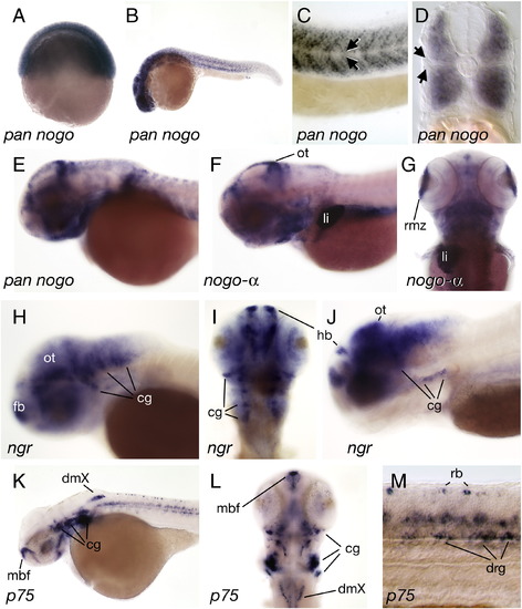
Expression of nogo, nogo receptor, and p75 in the developing zebrafish. (A) nogo transcripts were detected as early as 6 hpf. (nogo-α was not detected at this stage, not shown). (B) At 24 hpf, nogo was expressed in many neural and non-neural tissues but (C, D) expression was absent from a discrete domain (arrows) between the dorsal and ventral trunk muscles. (E, F, G) At 2 and 3 dpf, nogo transcripts were abundant in the posterior rim of the optic tectum (ot) and the marginal zone of the retina (rmz). Strong nogo-α expression was also detected in the liver starting at 3 dpf. (H) at 36 hpf and (I, J) 4 dpf ngr was expressed in many neural populations including cells in the forebrain (fb), habenula (hb), optic tectum, and cranial ganglia (gc). (K–M) Gene expression for the low-affinity neurotrophin receptor p75 was detected in the medial basal forebrain (mbf), cranial ganglia, dorsal motor nuclei of the vagus (dmX) and, at lower levels, in the retina at 2 dpf. (M) In the spinal cord, p75 was expressed in Rohon-Beard neurons (rb) and dorsal root ganglion neurons (drg). (A–C, E, F, H, J, K, M) side views, (D) cross section, (G, I, L) dorsal views. |
| Genes: | |
|---|---|
| Fish: | |
| Anatomical Terms: | |
| Stage Range: | Shield to Day 4 |
Reprinted from Molecular and cellular neurosciences, 40(4), Brösamle, C., and Halpern, M.E., Nogo-Nogo receptor signalling in PNS axon outgrowth and pathfinding, 401-409, Copyright (2009) with permission from Elsevier. Full text @ Mol. Cell Neurosci.