
Fig. 2
- ID
- ZDB-FIG-090304-2
- Publication
- Slanchev et al., 2009 - Control of Dead end localization and activity – Implications for the function of the protein in antagonizing miRNA function
- Other Figures
- All Figure Page
- Back to All Figure Page
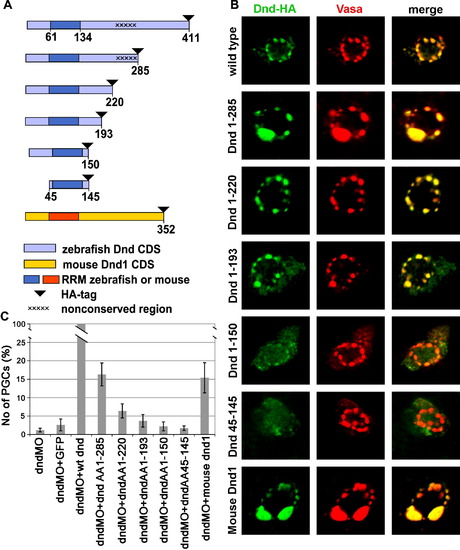
Deletions of parts of Dead end protein reveal domains important for protein localization and function. (A) Schematic drawing of Dead end deletion constructs. (B) Intracellular localization of the various Dnd-deletion fusion proteins detected by immunohistochemistry with anti-HA antibody (Green) in comparison with the position of the GCGs reported by anti-vasa antibody staining (Red). (C) A chart depicting the ability of the various deletions constructs to supplement for wild type Dead end. The values represent the number of rescued PGCs in embryos co-injected with a specific Dnd truncated protein and dnd MO, normalized to the PGCs rescued by a wild type dnd mRNA injection (see the text for details). |
| Fish: | |
|---|---|
| Knockdown Reagent: | |
| Observed In: | |
| Stage: | Unknown |
Reprinted from Mechanisms of Development, 126(3-4), Slanchev, K., Stebler, J., Goudarzi, M., Cojocaru, V., Weidinger, G., and Raz, E., Control of Dead end localization and activity – Implications for the function of the protein in antagonizing miRNA function, 270-277, Copyright (2009) with permission from Elsevier. Full text @ Mech. Dev.