
Fig. S2
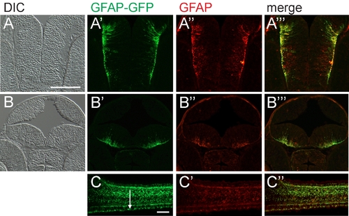
Green fluorescent protein (GFP) expression of embryonic tg(GFAP-GFP) closely resembles endogenous glial fibrillary acidic protein (GFAP). A,B: Transverse cryosections at two levels of the forebrain (differential interference contrast image). A′,B′: tg(GFAP-GFP) transgene expression. A″,B″: Immunohistochemistry with anti-GFAP antibody. A′″,B′″: Merged expression patterns of tg(GFAP-GFP) transgene and endogenous GFAP. C: Sagittal cryosection of the spinal cord of tg(GFAP-GFP). C′: Anti-GFAP antibody staining of the same section. C″: Merged panels showing overlap of the expression patterns of GFAP-GFP and endogenous GFAP. Expression is also detected in the floor plate (arrow). Anterior left and dorsal up. The 48 hours postfertilization (hpf) embryos are shown in A and B and the 42 hpf embryo is in C. Scale bar = 50 μm in A,C. |