
Fig. S5
- ID
- ZDB-FIG-090220-72
- Publication
- Carreira-Barbosa et al., 2009 - Flamingo regulates epiboly and convergence/extension movements through cell cohesive and signalling functions during zebrafish gastrulation
- Other Figures
- All Figure Page
- Back to All Figure Page
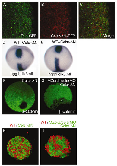
Characterisation of Celsr-ΔN. (A-C) Wild-type embryos were injected with 150 pg Dsh-GFP together with 100 pg Celsr-ΔN-RFP RNA. Scanning for GFP (A) and RFP (B) was carried out simultaneously and merged (C). Note that Celsr-ΔN is capable of recruiting Dsh without exogenous Fz, suggesting activation of the Wnt/PCP pathway. (D,E) Celsr-ΔN causes CE defects. Wild-type embryos were injected with 100 pg Celsr-ΔN-Venus RNA, and fixed at tail-bud for staining with hgg1 for the prechordal plate, ntl for the notochord and dlx3 for the anterior edge of the neural plate (see Table S1 in the supplementary material). (F,G) Effects of Celsr-ΔN on epiboly. Injection of a high dose (300 pg) Celsr-ΔN-Venus RNA does not lead to epiboly defects (F), whereas expression of 100 pg Celsr-ΔN-Venus RNA fails to rescue the celsr mutant/morphant phenotype (G; see Table S2 in the supplementary material). An arrowhead indicates the leading edge of deep cells. (H,I) Hanging drop assays. Cells from wild-type embryos with cells from embryos expressing 300 pg Celsr-ΔN RNA, being intermingled well (H) (n=11). Expression of 50 or 100 pg Celsr-ΔN-Venus RNA fails to restore altered cell cohesive property of celsr mutant/morphant cells (I) when compared with Fig 7C. Note that there is a strong correlation of epiboly defects with altered cell cohesive properties. |