
FIGURE
Fig. S2
Fig. S2
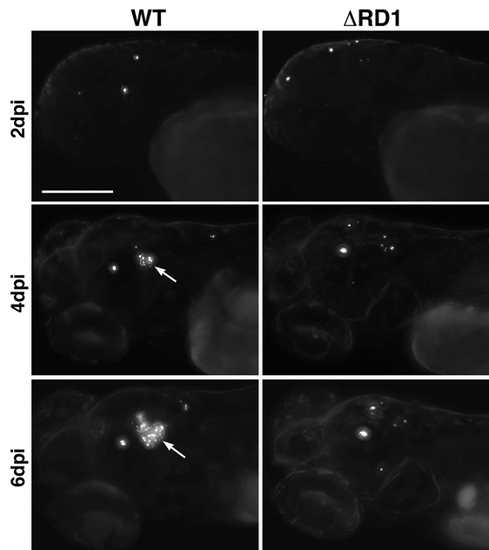
Overall progression of infection after hindbrain ventricle injection of 50-100 CFU of WT (left) or ΔRD1 (right) Mm. Though the burden of bacteria at 2dpi appears similar, the advent of a granuloma (arrows) in WT by 4dpi results in dramatically greater bacterial numbers by 6dpi. All bacteria present in these animals were only found in the head (data not shown), thus granuloma formation does not proceed from recruitment of infected cells. Scale bar, 200μm. |
Expression Data
Expression Detail
Antibody Labeling
Phenotype Data
Phenotype Detail
Acknowledgments
This image is the copyrighted work of the attributed author or publisher, and
ZFIN has permission only to display this image to its users.
Additional permissions should be obtained from the applicable author or publisher of the image.
Reprinted from Cell, 136(1), Davis, J.M., and Ramakrishnan, L., The role of the granuloma in expansion and dissemination of early tuberculous infection, 37-49, Copyright (2009) with permission from Elsevier. Full text @ Cell