|
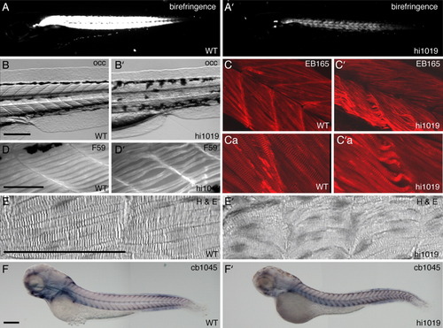
Fad24hi1019 mutants exhibit muscle degeneration. A-F: WT larvae. A′-F′: fad24hi1019 mutant larvae. A,A′: Polarized light microscopy to reveal birefringence of muscle. B,B′: OCC images of trunk muscle. C,C′: Confocal projections of trunk muscle immunolabeled with the fast-muscle myosin specific antibody EB165. Ca,C′a: Zoomed in images better showing muscle fiber striation and size. D,D′: Immunolabeling with the slow-muscle myosin specific antibody F59. E,E′: Hematoxylin and Eosin-stained longitudinal sections through trunk muscle tissue. F,F′: In situ hybridization using probe cb1045 to reveal myoseptum separating the somites of developing larvae. Lateral view, anterior to the left at 3 dpf. Scale bar = 200 μm. Representative results from three experiments with greater than 10 larvae per condition per experiment.
|