FIGURE

Fig. S2

ID
ZDB-FIG-081117-49
Publication
Bessarab et al., 2008 - Six1a is required for the onset of fast muscle differentiation in zebrafish
Other Figures
All Figure Page
Back to All Figure Page
Fig. S2

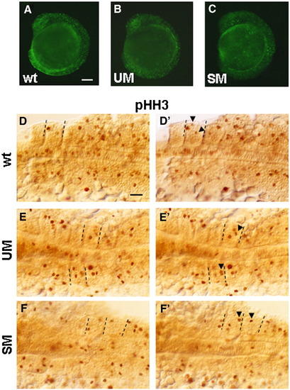
Staining with acridine orange and anti-phosphohistone H3 Ab at 9 s. (A–C) Fluorescent images of control and morphant embryos stained with acridine orange. Dorsal (D, E, F) and ventral (D′, E′, F′) focal planes of flat mounted embryos after immunohistochemistry with anti-phosphohistone H3 Ab. Posterior 6–7 somites are shown. Equal numbers (n = 10) of control embryos and morphants were stained. Arrowheads in D′, E′, F′ indicate phosphohistone H3-positive cells not clearly seen in D, E, F. Dashed lines mark the borders of somites containing these cells. Stained cells were counted in all 9 somites of 3 embryos for each condition (control, UM and SM morphants) and results were summarized in Table 1. No change in phoshohistone-positive cells in notochord was found. Scale bars, 100 μm.

Expression Data
Antibody:
Fish:
Knockdown Reagents:
Anatomical Terms:
Stage: 5-9 somites

Expression Detail
Antibody Labeling
Phenotype Data
Fish:
Knockdown Reagents:
Observed In:
Stage: 5-9 somites

Phenotype Detail
Acknowledgments
This image is the copyrighted work of the attributed author or publisher, and ZFIN has permission only to display this image to its users. Additional permissions should be obtained from the applicable author or publisher of the image.

Reprinted from Developmental Biology, 323(2), Bessarab, D.A., Chong, S.W., Srinivas, B.P., and Korzh, V., Six1a is required for the onset of fast muscle differentiation in zebrafish, 216-228, Copyright (2008) with permission from Elsevier. Full text @ Dev. Biol.