|
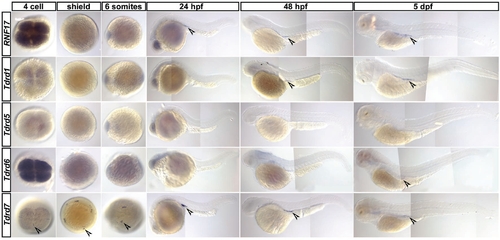
Expression pattern of Tudor domain containing proteins in zebrafish embryos and larvas. In situ hybridization for zebrafish homologs of previously described tudor domain containing genes RNF17, Tdrd1, Tdrd5, Tdrd6 and Tdrd7 at different stages starting from 4cell stage until 5 dpf. For RNF17 germ cell specific expression was observed starting from 24 hpf. For Tdrd1 germ cell specific expression was observed starting from 48 hpf. For Tdrd5 no expression could be observed in the analyzed stages. For Tdrd6 faint staining at the region where germ cells reside is visible at 3 dpf and 5 dpf. Tdrd7 expression is observed specifically at the cleavage furrow of the 4cell stage where the germ plasm is localized and continues to be expressed specifically in the germ cells throughout the analyzed stages.
|