
Fig. 4
- ID
- ZDB-FIG-080701-14
- Publication
- Strasser et al., 2008 - Control over the morphology and segregation of Zebrafish germ cell granules during embryonic development
- Other Figures
- All Figure Page
- Back to All Figure Page
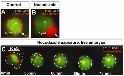
Granules structure is affected after microtubule disruption. A) Confocal section of germ cells in control embryos exposed to DMSO for 6 hours. Germ cell granules show normal perinuclear localization and distribution into small structures (arrow). B) Confocal section of germ cells in embryos exposed to 1 μg/ml of nocodazole for 6 hours. Nocodazole treated cells exhibit a conglomeration of Vasa positive granules forming a large granule (arrow). C) Embryos exposed to same conditions mentioned above. Vasa-dsRed labels granules, LaminB2-GFP labels nuclear envelop. The experiment shows how after laminB2 network disassembles, individual granules fuse forming bigger structures (asterisk). Cell outline is depicted with white dashed lines. All experiments were done in 11 hpf embryos. |