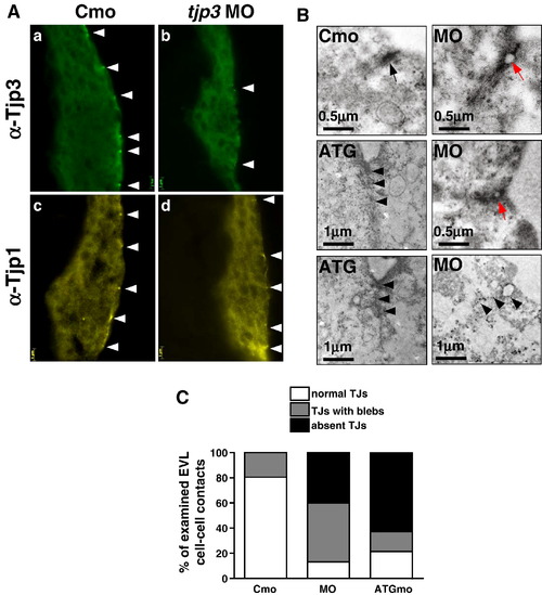
The loss of Tjp3/Zo-3 leads to a disruption of the TJ ultrastructure. (A) Cryosections of 6 hpf embryos stained with anti-Tjp3/Zo-3 (a, b) and anti-Tjp1/Zo-1 (c, d). Embryos were injected with control morpholino (a, c, Cmo) or tjp3/zo-3 splice morpholino (b, d, MO). Tjp3/Zo-3 protein is absent from most TJs in the EVL of morpholino injected embryos (b), but no phenotype is observed at that stage yet. The same embryos show a slight reduction in Tjp1/Zo-1 staining (d). (B) TEM of morpholino injected embryo sections show a marked loss and/or a disruption of the electron dense plaque of junctions in the EVL of morphants at 6 hpf. In control morpholino injected embryos, TJs can be observed at every cell–cell contact between neighboring cells of the EVL (Cmo, arrow). In the splice morpholino injected embryos, more than 40% of the EVL cell–cell contacts had discontinuities (blebbing) (MO, red arrows) and another 40% of contacts did not have any electron dense plaque (MO, arrowheads). Embryos injected with tjp3/zo-3 translation blocking morpholino had an even greater number of missing TJs (ATG, arrowheads). (C) Statistical analysis of TJ integrity as seen in TEM at 6 hpf. Two control and five morpholino injected embryos were analyzed. All contacts between adjacent EVL cells were classified into one of three groups. 1) Normal TJs, where cell membranes were in close contact throughout the length of the TJ, 2) discontinuous TJs with blebs, where cell membranes detached from each other, thus reducing the effective length of the TJ seal, and 3) junctions where no electron dense plaque was found between neighboring EVL cells. About 25% of TJs in control embryos had blebs (bar 1). In splice morpholino injected embryos, 47% of TJs showed blebbing and another 40% of the junctions were missing an electron dense plaque (bar 2). 62% of EVL cell–cell contacts in ATGmo injected embryos lacked an electron dense plaque (bar 3). N = 3 for Cmo, n = 3 for ATGmo, n = 5 for splice MO; number of cell–cell contacts counted per embryo 6–9; 2 independent experiments; t-test; p < 0.05.
|
