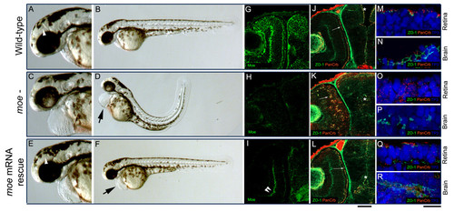
Injection of moe mRNA rescues defects in moe- embryos. (A-B) At 60 hpf, in wild-type embryos, the floor of the diencephalic ventricle is visible (A, white arrow head), the RPE is uniform, and the body axis is straight. (C, D) In moe- embryos, the ventricles are small or absent, the RPE is patchy, the tail curves and there is pericardial edema (D, arrow). (E, F) In moe- embryos injected with moe mRNA, the floor of the diencephalic ventricle is visible (E, white arrow head), the RPE is uniform, and the body axis is straight but mild pericardial edema persists (F, arrow). Anti-Moe labeling of 60 hpf wild-type embryos (G), moe- embryos (H), and moe- embryos injected with moe mRNA (I): the plexiform labeling in moe- embryos injected with moe mRNA (I, double arrowheads) is largely background. Adherens junctions (ZO-1, gnjected with moe mRNA (I): the plexiform labeling in moe- embryos injected with moe- embryos injected with moe mRNA (L), but are ectopically localized within the developing eye (arrow) and at the presumptive brain midline in moe- mutants with abnormal ventricle formation (asterix, K). High magnification confocal z-projections of TO-PRO-3 nuclear staining, and ZO-1 and panCrb labeling in the retina and brain in wild-type (M, N), moe- (O, P), and moe- embryos injected with moe mRNA (Q, R) at 60 hpf. (G, J, H, K, I and L are all single confocal z-sections). Scale bars, 50 μm (G-L), 10 μm (M-R).
|
