
Fig. 1
- ID
- ZDB-IMAGE-080402-16
- Genes
- Publication
- Christensen et al., 2008 - Tissue-specific requirements for specific domains in the FERM protein Moe/Epb4.1l5 during early zebrafish development
- All Figures
- Figures for Christensen et al., 2008
|
Fig. 1
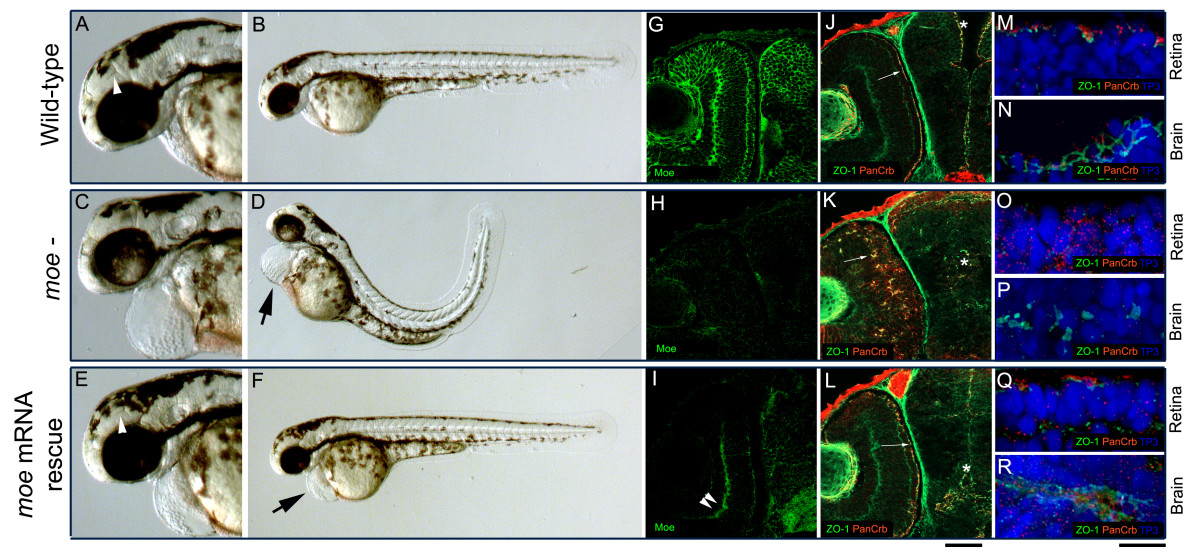
Injection of moe mRNA rescues defects in moe- embryos. (A-B) At 60 hpf, in wild-type embryos, the floor of the diencephalic ventricle is visible (A, white arrow head), the RPE is uniform, and the body axis is straight. (C, D) In moe- embryos, the ventricles are small or absent, the RPE is patchy, the tail curves and there is pericardial edema (D, arrow). (E, F) In moe- embryos injected with moe mRNA, the floor of the diencephalic ventricle is visible (E, white arrow head), the RPE is uniform, and the body axis is straight but mild pericardial edema persists (F, arrow). Anti-Moe labeling of 60 hpf wild-type embryos (G), moe- embryos (H), and moe- embryos injected with moe mRNA (I): the plexiform labeling in moe- embryos injected with moe mRNA (I, double arrowheads) is largely background. Adherens junctions (ZO-1, gnjected with moe mRNA (I): the plexiform labeling in moe- embryos injected with