
Fig. 7
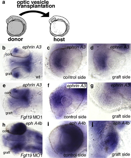
Nasal–temporal patterning of the retina after heterotopic optic vesicle transplantation. (a) Transplantation scheme: optic vesicles were grafted from wild-type donors to wild-type hosts, and from Fgf19 MO1-injected donors to wild-type hosts at the 10-somite stage. (b–j) Nasal–temporal patterning of the retina after transplantation at 28 h of development. (b–d) In optic vesicle grafts from wild-type embryos, the expression of ephrin A3 was correctly detected in the nasal retina. (e–g) In optic vesicle grafts from Fgf19 MO1-injected donors, the expression of ephrin A3 was lost in the nasal half. (h–j) In optic vesicle grafts from Fgf19 MO1-injected donors, the expression of eph A4b was slightly expanded into the nasal retina. (b, e, h) Dorsal views with anterior to the left. (c, d, f, g, i, j) Lateral views with anterior to the left and dorsal to the top. |
Reprinted from Developmental Biology, 313(2), Nakayama, Y., Miyake, A., Nakagawa, Y., Mido, T., Yoshikawa, M., Konishi, M., and Itoh, N., Fgf19 is required for zebrafish lens and retina development, 752-766, Copyright (2008) with permission from Elsevier. Full text @ Dev. Biol.