
Fig. 5
- ID
- ZDB-FIG-080326-66
- Publication
- Pietri et al., 2008 - Six cadm/synCAM genes are expressed in the nervous system of developing zebrafish
- Other Figures
- All Figure Page
- Back to All Figure Page
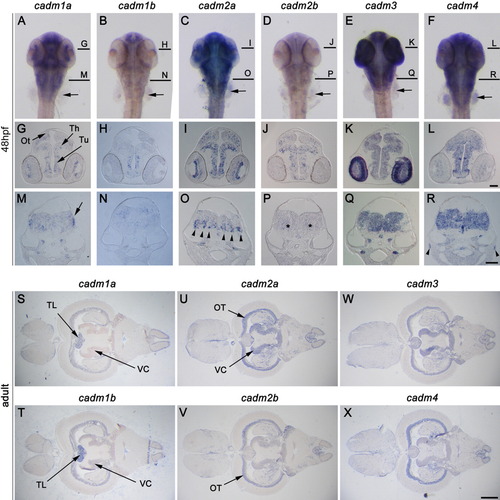
Expression of cadms in the brain. A-R: In situ hybridization (ISH) staining in 48 hours postfertilization (hpf) zebrafish embryos; a dorsal view of whole-mount zebrafish (A-F), and cross-sections at the midbrain (G-L), and hindbrain (M-R) are presented for each cadm gene. Lines in A-F represent levels of the sections in G-L and M-R. S-X: cadm expression visualized in horizontal sections of adult brain. The cadm genes are expressed broadly in the developing and adult brain and show partially overlapping domains of expression. cadm2a is strongly expressed in the ventral medulla oblongata (MO) (arrowheads in O), while cadm2b is more diffuse in this region (asterisks in P). The posterior part of the MO shows strong staining for cadm1a (arrow in M). Ot, developing optic tectum; OT, optic tectum granular layer L3; Th, thalamus; TL, torus longitudinalis; Tu, tuberculum; VC, valvula cerebellis. Orientations are anterior at the top in A-F; dorsal at the top in G-R; anterior to the left in S-X. Scale bars = 50 μm in G-L, 50 μm in M-R, 0.5 mm in S-X. |