|
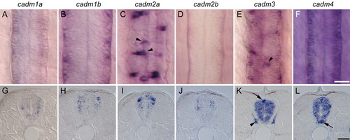
Expression of cadms in the developing spinal cord. A-L: The cadm expression revealed by in situ hybridization in whole-mount (dorsal view, A-F) and cross-sections (G-L) of 48 hpf embryos. Due to the strong rostrocaudal gradient of cadm1b expression, panels B and H show an anterior localized region of the spinal cord (somites 3-5), immediately behind the hindbrain, whereas all other images are from a region dorsal to the anus (somites 12-15). Staining for cadm2a and 3 is in dorsal cells at the midline of the spinal cord, suggestive of sensory Rohon-Beard neurons (arrowheads in C and E, arrow in K). Expression for cadm3 is also seen in the dorsal root ganglion (arrowhead in K). cadm4 is strongly expressed in the ventral domain of the spinal cord indicative of floor plate or motoneurons. Scale bars = 20 μm in A-F, 20 μm in G-L.
|