|
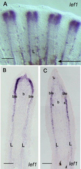
Comparison of the expression of lef1 in a 4-day fin regenerate using the two ISH methods. A: Whole-mount ISH with a lef1 probe on 4-dpa regenerates shows expression splitting into two domains in the distal part of each fin ray indicating an imminent bifurcation event. B: Upon cryo-sectioning of the fin regenerate shown in A, lef1 expression is observed in the basal layer of the epidermis and in the distal blastema. C: A similar expression pattern is observed when an ISH is performed on cryo-sections of 4-dpa regenerates. The arrow in A indicates the level of amputation. L, lepidotrichia; b, blastema; ble, basal layer of epidermis; s, scleroblasts. Scale bars = 100 mu;m (A), 50 μm (B,C).
|