
Fig. 1
- ID
- ZDB-FIG-080324-5
- Publication
- Smith et al., 2008 - Gene expression analysis on sections of zebrafish regenerating fins reveals limitations in the whole-mount in situ hybridization method
- Other Figures
- All Figure Page
- Back to All Figure Page
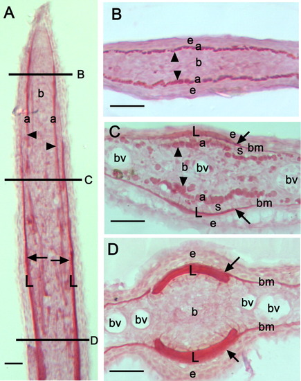
Picrosirius red staining of collagen on sections of 4-day regenerates. Longitudinal (A) and transverse (B-D) cryo-sections of a 4 dpa regenerated fin ray that has been stained with picrosirius red, which labels collagenous structures, including the lepidotrichia (arrows), the actinotrichia (arrowheads) and the basement membrane. The horizontal lines in A indicate the approximate positions of the transverse sections presented in B-D along the proximal-distal axis of the regenerate. The level of amputation of the fin is not shown in A. Note the presence of the tip of the actinotrichia in the distal part of the regenerate (B). More proximally, both actinotrichia of increased diameter and the lepidotrichia bone matrix are visible (C), while in the proximal part of the regenerate only thick lepidotrichia are present (D). L, lepidotrichia; a, actinotrichia; b, blastema; e, epidermis; bm, basement membrane; bv, blood vessels; s, scleroblasts. Scale bars = 25 μm (A-D). |