|
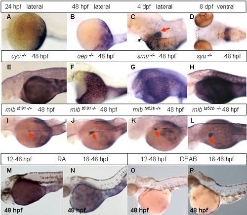
Analyses of rbp4 expression during late development and its regulation. (A-D) rbp4 expression in 24 hpf, 48 hpf, 4 dpf and 8 dpf wild-type embryos as indicated. A red arrow indicates the liver while a black arrow indicates the pericardium region. (E-H) rbp4 expression in cyc-/-, oep-/-, smu-/- and syu-/- embryos at 48 hpf as indicated. (I – L) rbp4 expression in various heterozygous and homozygous mib mutant embryos as indicated. Red arrows indicate the liver. Note precocious appearance of rbp4 expression in homozygous mutant liver at 48 hpf (J, L) compared to heterozygous mib embryos (I, K). (M, N) rbp4 expression in 48 hpf embryos treated with 10-6 M RA initiated from 12 hpf (M) or 18 hpf (N). (O, P) rbp4 expression in 48 hpf embryos treated with 10-5 M DEAB initiated from 12 hpf (O) and 18 hpf (P).
|