
Fig. 2
- ID
- ZDB-FIG-080225-21
- Publication
- Patterson et al., 2008 - Growth in the larval zebrafish pectoral fin and trunk musculature
- Other Figures
- All Figure Page
- Back to All Figure Page
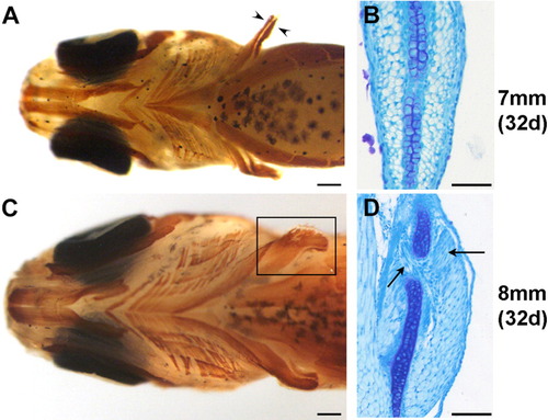
Changes in pectoral fin musculature between 7.0 mm standard length (SL; A,B) and 8.0 mm SL (C,D), A: Ventral view of the musculature of the 7.0 mm larva. Musculature has been labeled in whole-mount with MF20 antibody (brown). The pectoral fin musculature consists of two distinct masses on either side of the endoskeletal disk (arrowheads). B: Transverse section of the pectoral fin of a 7.0 mm larva stained with methylene blue, showing muscle masses on either side of the central endoskeletal disk. C: Ventral view of the musculature of the 8.0 mm larva labeled with MF20 (brown); the fin muscle is boxed. D: Transverse section of a pectoral fin of an 8.0 mm larva demonstrating the beginning of splitting of the muscle masses (arrows). Scale bars 250 μm in A,C, 50 μm in B,D. |