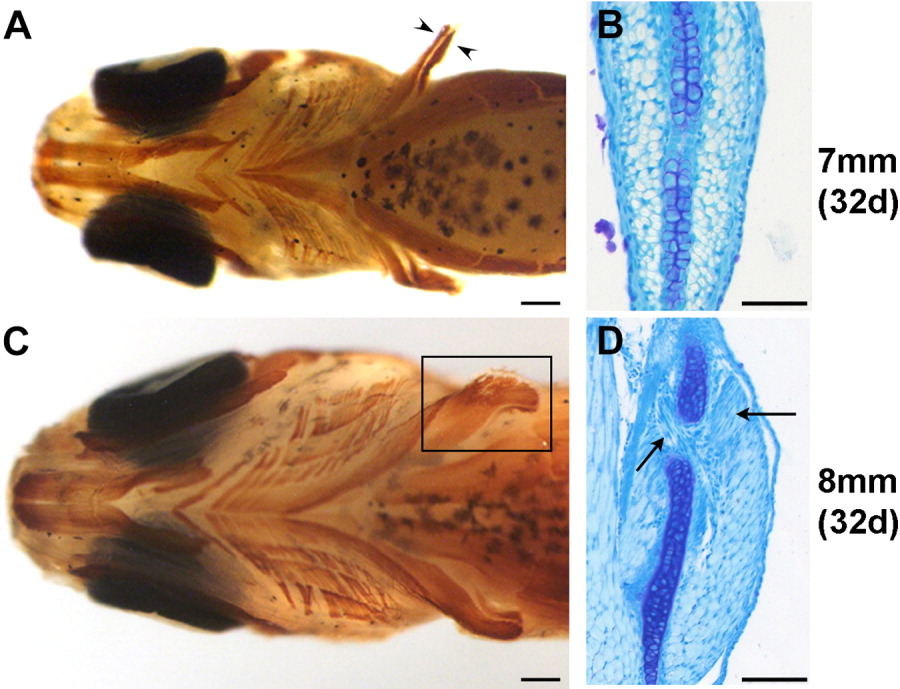
Fig. 2 Changes in pectoral fin musculature between 7.0 mm standard length (SL; A,B) and 8.0 mm SL (C,D), A: Ventral view of the musculature of the 7.0 mm larva. Musculature has been labeled in whole-mount with MF20 antibody (brown). The pectoral fin musculature consists of two distinct masses on either side of the endoskeletal disk (arrowheads). B: Transverse section of the pectoral fin of a 7.0 mm larva stained with methylene blue, showing muscle masses on either side of the central endoskeletal disk. C: Ventral view of the musculature of the 8.0 mm larva labeled with MF20 (brown); the fin muscle is boxed. D: Transverse section of a pectoral fin of an 8.0 mm larva demonstrating the beginning of splitting of the muscle masses (arrows). Scale bars 250 μm in A,C, 50 μm in B,D.
Image
Figure Caption
Acknowledgments
This image is the copyrighted work of the attributed author or publisher, and
ZFIN has permission only to display this image to its users.
Additional permissions should be obtained from the applicable author or publisher of the image.
Full text @ Dev. Dyn.

