
Fig. 2
- ID
- ZDB-FIG-071213-14
- Publication
- Murciano et al., 2007 - Position dependence of hemiray morphogenesis during tail fin regeneration in Danio rerio
- Other Figures
- All Figure Page
- Back to All Figure Page
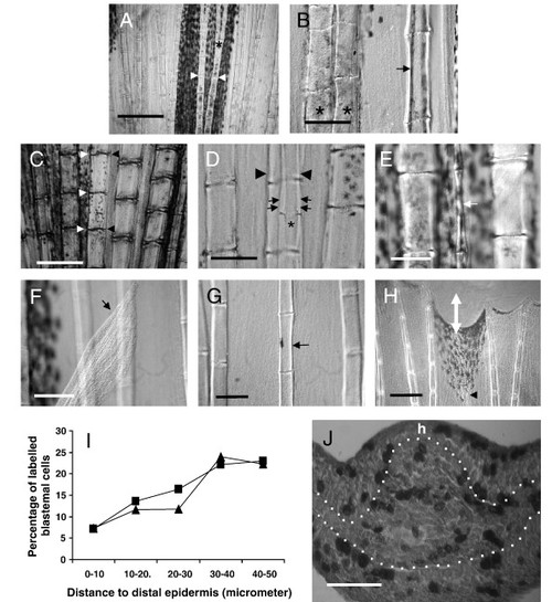
The hemiray, or a hemiray fragment, may regenerate in the caudal fin of Danio rerio. (A) The hemirays of a given ray are symmetrical. Segment joints (arrowheads) and bifurcations (asterisk) occur in exactly the same place in both hemirays. (B) After operation A, the hemiray regenerates showing a much thinner profile (arrow) and do not bifurcate. Compare the width of the regenerated hemiray (arrow) with the neighboring ray (left). Control ray regenerate normally bifurcate (asterisk) whereas hemirays regenerates do not. (C) After operation B, a thin H3 regenerate (small arrows) is obtained. Loss of coordination between segment joints occurs between regenerating hemiray (asterisk) and contralateral extant hemiray (arrowheads). (D) After operation B, partial hemiregenerates (white arrow) can also be obtained. Hemiray width was extremely narrow in this case and the complete original size was not achieved. The lengths of the segments, however, are similar to those in the neighboring rays. (E) After operation C, the hemiray may regenerate outside of the fin (arrow). In these cases, the size of the neighboring rays was never achieved after 1 month. (F) A detail of an H1 regenerate growing inside the fin after operation C. The width of the hemiray (arrow) is thinner than control neighboring rays. (G) A detail of distal regions of a regenerate after cut following operation C. Observe the hemiray (arrowhead) nearly reaches the complete size of neighboring rays (white double arrow shows non-regenerated distance). (H) Graphic showing the average percentage of BrdU-labeled blastemal cells against distance to distal epidermis (μm). ▲: hemiray blastemas, •: ray blastemas. Observe that distal 10 μm blastemal cells rarely incorporate BrdU in both hemiray and ray blastema. (I) Transversal section of a proliferating hemiblastema after operation A. BrdU is incorporated to dividing cells in both blastemal mesenchyme and epidermis. h shows the side in which the hemiray regenerate. Scale bar represents 500 μm (A), 300 μm (G), 125 μm (C, F), 100 μm (B, D, E) and 50 μm (I). |
| Genes: | |
|---|---|
| Fish: | |
| Condition: | |
| Anatomical Term: | |
| Stage: | Adult |
Reprinted from Developmental Biology, 312(1), Murciano, C., Pérez-Claros, J., Smith, A., Avaron, F., Fernández, T.D., Durán, I., Ruiz-Sánchez, J., García, F., Becerra, J., Akimenko, M.A., and Marí-Beffa, M., Position dependence of hemiray morphogenesis during tail fin regeneration in Danio rerio, 272-283, Copyright (2007) with permission from Elsevier. Full text @ Dev. Biol.