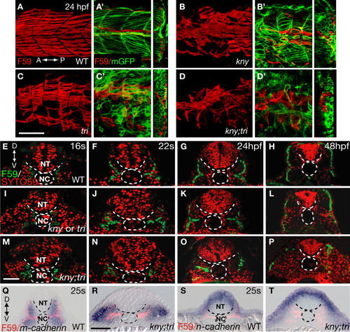
kny;tri double mutants are defective in slow muscle migration. A-D: Lateral views of the trunk region of the embryos stained with the slow muscle marker F59 antibody. Confocal z-stacks were collected and merged into one focal plane. A'-D': Left panels show the lateral views of the same embryos as shown in A-D, but only one single focal plane of the z-stacks is presented. The embryos are labeled with mGFP to highlight the morphology of the myotome and individual muscle fibers. Right panels show the confocal 3D reconstructed transverse sections. E-P: Localization of the slow muscle cells between the 16-somite stage (17 hpf) and 48 hpf as revealed by F59 antibody staining. SYTO59, which labels the nuclei, shows the embryonic structures. Q-T: m- and n-cadherin RNA expression (blue) and the positions of slow muscle cells revealed by F59 antibody labeling (pink) at the 25-somite stage (21.5 hpf). E-T: Transverse sections of the second or third somite. A, anterior; D, dorsal; NC, notochord; NT, neural tube; P, posterior; V, ventral. Scale bars = (A-D, A'-D', Q-T) 50 μm, (E-P) 20 μm.
|
