|
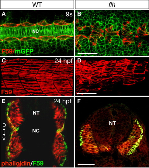
The presence of an intact notochord is not essential for slow muscle morphogenesis. A, B: Morphology of the adaxial cells in WT (A) and flh (B) mutant embryos at the 9-somite stage (13.5 hpf) as revealed by mGFP and F59 labeling. Notice the absence of the notochord and the aberrant positions of the adaxial cells in flh mutants (B). Dorsal views, anterior to the left. C,D: Confocal images of the slow muscle fibers stained with F59 antibody in WT (C) and flh mutants (D). Lateral views, anterior to the left. E,F: Transverse sections of the anterior myotomes stained with F59 antibody that labels the slow muscle cells, and phalloidin that recognizes both the fast and slow muscle fibers (Henry and Amacher,[2004]). A, anterior; D, dorsal; P, posterior; NT, neural tube; NC, notochord; V, ventral. Scale bars = 50 μm in A-F.
|