
Fig. 5
- ID
- ZDB-FIG-071019-11
- Publication
- Yin et al., 2007 - Convergence and extension movements affect dynamic notochord-somite interactions essential for zebrafish slow muscle morphogenesis
- Other Figures
- All Figure Page
- Back to All Figure Page
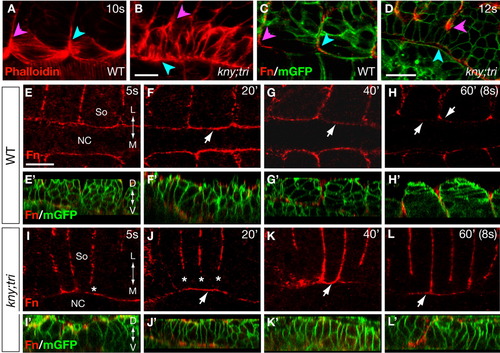
The adaxial cells in kny;tri double mutants exhibit prolonged contact with the notochord. A, B: F-actin distribution at the 10-somite stage (14 hpf) as revealed by phalloidin staining. C, D: Expression of Fn protein at the 12-somite stage (15 hpf). The embryos were co-labeled with mGFP to illustrate the morphology of the adaxial cells and the notochord. In A-D, the pink arrowheads point to the positions where the adaxial cells contact the anterior somitic boundary, whereas the blue arrowheads point to the opposite end of the adaxial cells. E-L: Time-course analyses of the Fn protein expression between the 5- and 8-somite stages. Arrows in F-H and J-K point to the expression of Fn protein at the notochord surface during the adaxial cell shape changes. E'-L': Confocal reconstructed sagital sections of the same embryos as shown in E-L, but were co-labeled with mGFP and Fn antibody. A-L: Dorsal views, anterior to the left. L, lateral; D, dorsal; M, medial; NC, notochord; So, somite; V, ventral. Scale bars = 20 μm (A-L, E'-L'). |