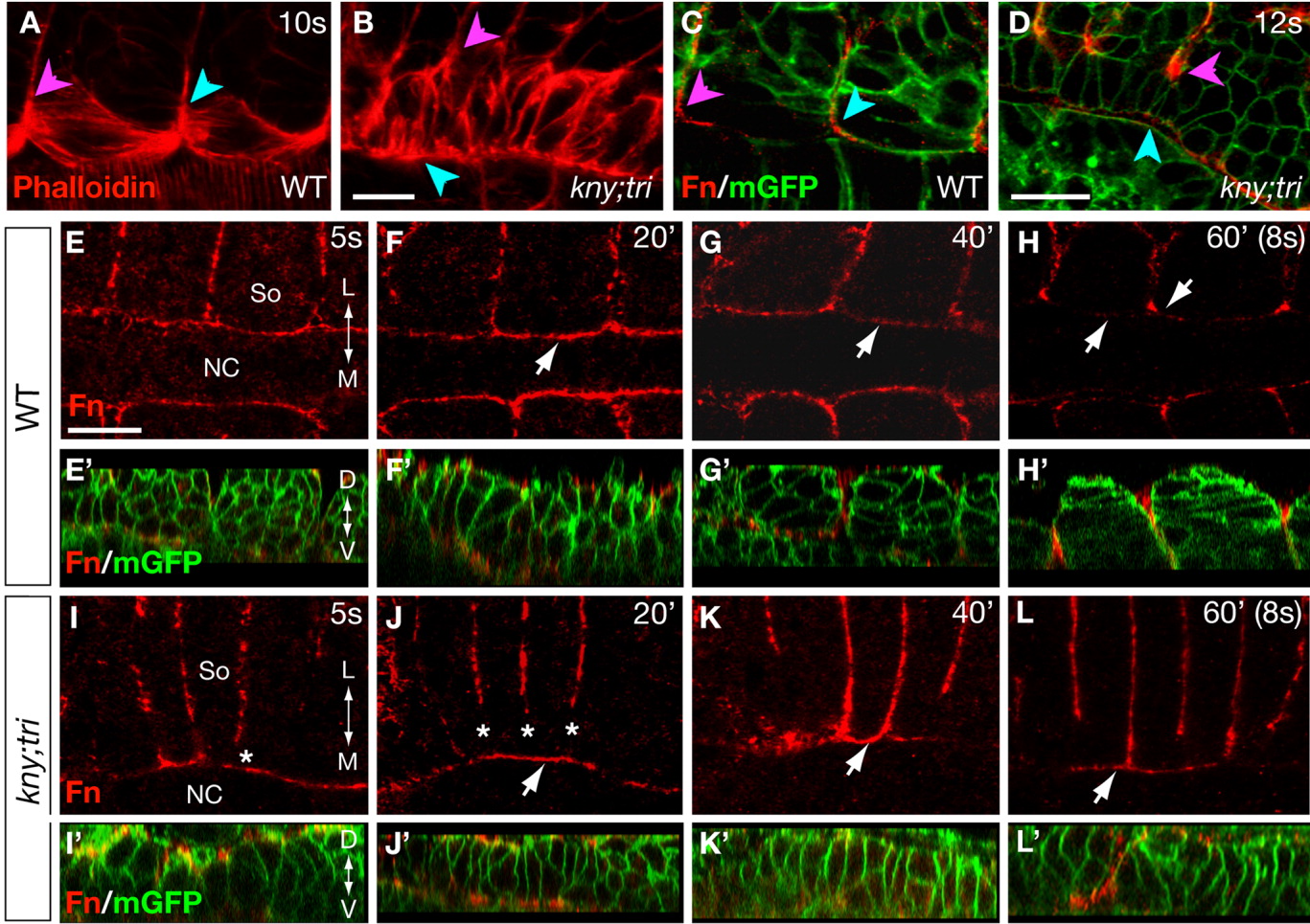
Fig. 5 The adaxial cells in kny;tri double mutants exhibit prolonged contact with the notochord. A, B: F-actin distribution at the 10-somite stage (14 hpf) as revealed by phalloidin staining. C, D: Expression of Fn protein at the 12-somite stage (15 hpf). The embryos were co-labeled with mGFP to illustrate the morphology of the adaxial cells and the notochord. In A-D, the pink arrowheads point to the positions where the adaxial cells contact the anterior somitic boundary, whereas the blue arrowheads point to the opposite end of the adaxial cells. E-L: Time-course analyses of the Fn protein expression between the 5- and 8-somite stages. Arrows in F-H and J-K point to the expression of Fn protein at the notochord surface during the adaxial cell shape changes. E'-L': Confocal reconstructed sagital sections of the same embryos as shown in E-L, but were co-labeled with mGFP and Fn antibody. A-L: Dorsal views, anterior to the left. L, lateral; D, dorsal; M, medial; NC, notochord; So, somite; V, ventral. Scale bars = 20 μm (A-L, E'-L').
Image
Figure Caption
Acknowledgments
This image is the copyrighted work of the attributed author or publisher, and
ZFIN has permission only to display this image to its users.
Additional permissions should be obtained from the applicable author or publisher of the image.
Full text @ Dev. Dyn.

