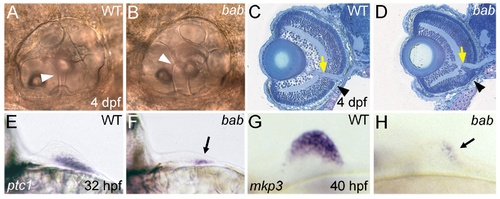
REREa is necessary for the proper development of multiple tissue types. (A,B) Lateral views with anterior to the left of otic capsules (ears) in live larvae at 4 dpf. The epithelial pillars (white arrowheads) that help to form the semicircular canals of the otic capsule fail to fuse in bab (B) compared to wild-type (A) larvae. (C,D) Methylene blue/azure II stains of frontal plastic sections through the head of wild-type (C) and bab (D) 4 dpf larvae. The yellow arrows mark where retinal tissue has merged with the optic nerve, while the black arrowheads mark disrupted retinal lamination in the ventral retina of bab compared to wild-type larvae. Note the compacted appearance of the bab retina compared to wild-type. (E-H) Views on the fin buds of whole-mount in situ hybridizations of wild-type (E,G) and bab (F,H). (E,F) Frontal view with medial to the left of ptc1 expression at 32 hpf. (G,H) Lateral view with anterior to the left of mkp3 expression at 40 hpf. Arrows in (F) and (H) denote the residual fin bud expression of ptc1 and mkp3, respectively. In both cases (ptc1 and mkp3), expression of these genes is initiated but not maintained in the fin bud.
|
