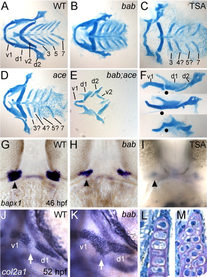
rerea genetically interacts with fgf8 in pharyngeal arch development, and disruption of histone deacetylase (HDAC) activity results in bab-like fusions of the first arch. A-M: Alcian blue-stained cartilage flat-mounts at 5 dpf (A-F), whole-mount in situ hybridizations of embryos with bapx1 (G-I) and col2a1 (J,K), and frontal sections through the palatoquadrate (d1 element, L,M) are shown for wild-type (WT; A,F top, G,J,L), bab (B,F middle, H,K,M), Trichostatin A (TSA) -treated (C,F bottom, I), ace (D), and bab;ace (E) embryos or larvae. Anterior is to the left in all images except G-I,L,M, which are frontal views on the mouth. A-E: Ventral views of flat-mounted cartilages with posterior arches numbered 3-7, reveal the presence but reduction in all elements in bab (B), a missing posterior arch in both TSA-treated (C) and ace (D), and a complete loss of posterior arches in bab;ace (E). Disruption of REREa (H) or TSA treatment (I) results in reduction of joint specification marker, bapx1 compared with wild-type embryos (G). Later col2a1 is misexpressed in cells of the presumptive joint (white arrow) in bab (K) compared with WT (J) leading to a fusion of the first arch cartilages (F, lateral view of arches 1 and 2) in wild-type (top F), bab (middle F), and TSA-treated (bottom F) embryos. Dots mark the position of the joint in each preparation. Chondrocyte morphology at 4 hpf is round in bab (M) compared with the stacked structure in wild-types (L). d1 and d2, dorsal cartilages of arches 1 and 2, respectively; v1 and v2, ventral cartilages of arches 1 and 2, respectively.
|
