
FIGURE
Fig. 3
Fig. 3
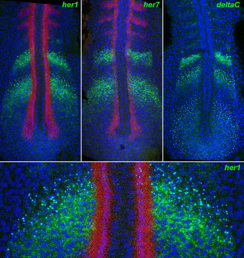
Nascent and Mature Transcripts Visualized by In Situ Hybridisation. Fluorescent staining by ISH for her1 (top left), her7 (top middle), and deltaC (top right), using tyramide chemistry (green), reveals nuclear dots corresponding to nascent transcripts and cytoplasmic signal corresponding to mature mRNA. The bottom panel is a magnified detail of the top left panel. The images are confocal optical sections of flat-mounted specimens, counterstained for DNA with TOPRO3 (blue false colour). Red staining shows myoD expression by dual ISH. |
Expression Data
| Genes: | |
|---|---|
| Fish: | |
| Anatomical Terms: | |
| Stage: | 5-9 somites |
Expression Detail
Antibody Labeling
Phenotype Data
Phenotype Detail
Acknowledgments
This image is the copyrighted work of the attributed author or publisher, and
ZFIN has permission only to display this image to its users.
Additional permissions should be obtained from the applicable author or publisher of the image.
Full text @ PLoS Biol.