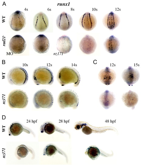
runx1 expression fails to initiate in the ALM and PLM of zebrafish embryos compromised for Rad21, but late neuronal runx1 expression is Rad21-independent. (A-D) Whole-mount embryos stained for runx1 expression. For all, upper panels are wild type, lower panels are rad21ATGMO morphants (MO) or nz171 homozygotes as indicated. (A) Posterior views of embryos, stages as indicated. rad21 morphants (MO; 0.5 pmol) at the 4-somite stage (4s; 14/15 embryos) and 6s (23/25 embryos) were runx1 negative. Expression of runx1 in RB cells is just detectable by the 8-somite stage in nz171 mutants, but PLM expression remains absent throughout. (B) Lateral views, stages as indicated. runx1 mRNA is not detected in the PLM or ICM, or in the olfactory placode of nz171 mutants, but is present in a population of RB cells. (C) Anterior views, stages as indicated. The occasional runx1-positive cell is visible in the ALM of nz171 mutants. (D) Lateral views, stages as indicated. At 24 h.p.f., nz171 homozygotes appear delayed and display no runx1 expression. By 28 h.p.f., neuronal and olfactory placode expression has initiated in mutants. Hematopoietic expression remains absent, c.f. wild type, where expression of runx1 is observed in the ventral wall of the dorsal aorta. Anterior is to the left for B and D.
|
