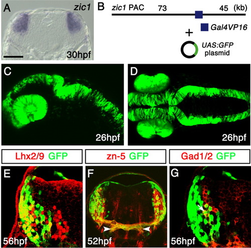
Green fluorescent protein (GFP) expression in the Tg(zic1:Gal4VP16/UAS:GFP) embryos. A: In situ hybridization of a 30-hours postfertilization (hpf) coronal section of the hindbrain at the level of the otic vesicle, using the zic1 probe. Dorsal is to the top. B: The structure of the DNA constructs used to generate the Tg(zic1:Gal4VP16/UAS:GFP) fish. The zic1 PAC with Gal4VP16 inserted into the 5′-untranslater region (5′-UTR) of zic1 was coinjected with the UAS:GFP plasmid. C,D: Lateral (C) and dorsal (D) views of the Tg(zic1:Gal4VP16/UAS:GFP) embryos at 26 hpf. GFP is expressed in the eyes and dorsal neural tube. E-G: Cross-sections of the hindbrain of the Tg(zic1:Gal4VP16/UAS:GFP) embryos stained with the Lhx2/9 (E), zn-5 (F), and Gad1/2 (G) antibodies. The lateral GFP(+) cells predominantly coexpressed Lhx2/9 (E) and the zn-5 antigen (F), while some of the medial GFP(+) cells coexpressed Gad1/2 (G). A representative double-positive cell is indicated with an arrow in G. (F) The axons of the GFP(+) neurons colocalized with the zn-5(+) commissural axons. These axons projected ventrolaterally after crossing the midline (arrowheads). They were connected with the longitudinal fascicles as shown in Figure 4B. UAS, upstream activating sequences. Scale bars = 60 μm in A, 100 μm in C,D, 25 μm in E,G, 50 μm in F.
|
