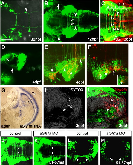
The zn-5(+)Lhx2/9(+) neurons project to the contralateral torus semicircularis (TS). A: Dorsal view of the hindbrain of a Tg(zic1:Gal4VP16/UAS:GFP) embryo at 30 hours postfertilization (hpf). The axon of a single green fluorescent protein-positive (GFP(+)) neuron can be seen turning rostrally after crossing the midline (arrowhead). Rostral is to the left. B: By 72 hpf, the GFP(+) axons formed a ladder-like structure, which consisted of many commissures and two longitudinal fascicles (filled arrowheads). The axon terminals of the two longitudinal fascicles appear to reach the lateral midbrain (arrows). Additionally, a pair of GFP(+) patches (open arrowheads) observed in rhombomere 4 (r4) around the initial axon segment of the Mauthner cell (M, compare with C) were probably Mauthner axon caps formed by the spiral fiber neurons and/or passive hyperpolarizing potential neurons (Lorent et al.,[2001]). The position of cell bodies providing the GFP(+) Mauthner axon caps was unclear. C: Reticulospinal neurons were visualized in the 3 days postfertilization (dpf) Tg(zic1:Gal4VP16/UAS:GFP) embryos by retrograde incorporation of tetramethylrhodamine-dextran. As in B, the longitudinal fascicles and Mauthner axon caps are indicated by the filled and open arrowheads, respectively. The majority of commissures were distributed from r2 to r6, and the caudal end of the longitudinal fascicle corresponded approximately to the caudal r6 border. Note the gross overlap of the GFP(+) neurons with the spreading of central nerves of the posterior lateral line (PLLn, arrow), which was also accidentally labeled by tetramethylrhodamine-dextran. M, Mauthner cell. D: Lateral view of the 4-dpf Tg(zic1:Gal4VP16/UAS:GFP) embryos. Anterior is to the left. The GFP(+) longitudinal fascicles appeared to terminate in the lateral midbrain region ventral to the optic tectum (arrow, compare with B). Tc, optic tectum. E,F: Dorsal views of a Tg(zic1:Gal4VP16/UAS:GFP) embryo with 1,1′, di-octadecyl-3,3,3′,3′,-tetramethylindo-carbocyanine perchlorate (DiI) applied on the contralateral dorsal hindbrain. Anterior is to the left. Images in E,F were obtained from the same embryo but at different dorsoventral focal planes. The image in E was obtained from the deeper (ventral) region, while the image in F was from the more superficial (dorsal) region. After crossing the midline, the DiI-labeled axons were segregated into the dorsal and ventral fascicles. E: The ventral fascicle turned rostrally, joined the anteriorly projecting longitudinal fascicle (filled arrowheads) and terminated in the GFP(+) region of the midbrain (arrow, compare with B). The posterior segment of the longitudinal fascicle (right filled arrowhead) derives from the anterior projection of caudal commissural axons, which are weakly labeled with the dye. F: The dorsal fascicle advanced straight, then bifurcated and terminated in the contralateral hindbrain with extensive branches (open arrows in E,F). Retrograde labeling of a cell body (asterisks) was also observed. A confocal section showed that the neuron was GFP(+) (inset). G: In situ hybridization of a coronal section of the adult midbrain, using the lhx9 probe. Dorsal is to the top, lateral is to the right. The expression of lhx9 is evident in the torus semicircularis (TS) and periventricular gray zone of the optic tectum (Tc). H,I: Immunostaining of a coronal section of the embryonic Tg(zic1:Gal4VP16/UAS:GFP) midbrain with the Lhx2/9 antibody. Cell nuclei (gray) are counterstained by SYTOX dye. The axon terminals of the GFP(+) longitudinal fascicles (arrow) were distributed through the entire mediolateral extent of the region expressing Lhx2/9. This staining pattern of Lhx2/9 in the embryo is essentially identical to that of lhx9 in adult fish (G), suggesting that the GFP(+) longitudinal fascicles formed by the zn-5(+)Lhx2/9(+) neurons project to the developing midbrain TS. J-M: Knockdown of Atoh1a impairs the formation of the lateral longitudinal fascicles (LLFs). J,K: Dorsal views of the control (J) and atoh1a MO-injected (K) Tg(zic1:Gal4VP16/UAS:GFP) embryos. In the atoh1a MO-injected embryos (K), the commissural fascicles are thin and LLFs (compare arrowheads in J,K) are absent. Anterior is to the left. L,M: Coronal sections of the hindbrain of the control (L) and atoh1a MO-injected (M) Tg(zic1:Gal4VP16/UAS:GFP) embryos. Dorsal is to the top. Commissural fascicles are very thin and LLFs are absent (compare arrowheads in L,M). UAS, upstream activating sequences. Scale bars = 50 μm in A, 100 μm in B, 75 μm in C, 100 μm in D, 50 μm in E,F,H,I, 200 μm in G, 80 μm in J,K, 60 m in L,M.
|
