
Fig. S1
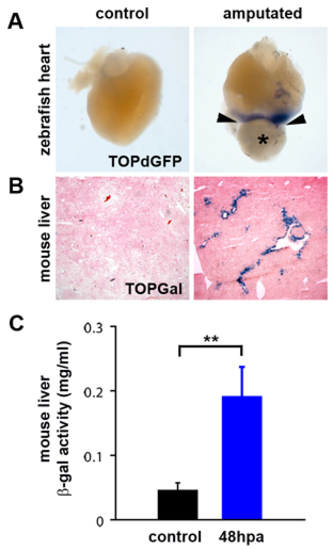
Wnt/β-catenin signaling is upregulated in regenerating zebrafish heart and mouse liver. (A) Wnt/β-catenin reporter (TOPdGFP) activity (detected by in situ hybridization for GFP RNA, blue) is upregulated in regenerating zebrafish hearts (n=4) adjacent to the amputation plane (arrowheads) at 3 dpa (*=blood clot). Control is a sham-operated TOPdGFP heart at 3 days post-sham. TOPdGFP is also expressed adjacent to the amputation plane at 7 dpa (n=3) and 14 dpa (n=3) (data not shown). (B) Wnt/β-catenin reporter (TOPGal) (DasGupta and Fuchs, 1999) activity (detected by staining for β-galactosidase activity, blue) is upregulated in periportal hepatocytes in regenerating mouse liver (n=8), 48 hours post-partial hepatectomy. (C) Quantification of TOPGal activity in regenerating mouse liver. β-galactosidase activity was measured in control lobes (resected lobes removed at the time of partial hepatectomy) and regenerating lobes, collected 48 hpa. Control lobes were compared to regenerating lobes harvested from the same animal. Asterisks indicate a statistically significant difference between experimental groups (n=8, P<0.01, Paired Student’s t-Test). Error bars represent the s.e.m. |