
Fig. 1
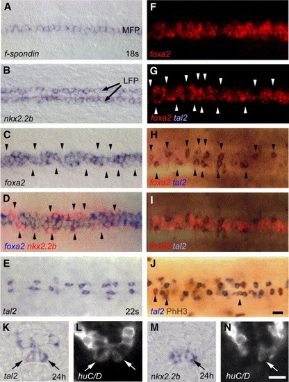
LFP cells show different expression profiles and identities. (A, B) Dorsal views of trunk regions showing homogenous expression of f-spondin in the MFP (A) and nkx2.2b in the LFP (B). (C) foxa2 is discontinuously expressed in the LFP, with regions of weak expression levels along the AP axis (indicated by arrowheads). (D) Overlay of a double in situ hybridization showing nkx2.2b (in red) that is strongly expressed within regions of weak foxa2 expression (in blue). In overlapping regions, nkx2.2b detection is quenched. (E) tal2 is expressed in single cells of the LFP that are discontinuously distributed along the AP axis. (F–I) Double in situ hybridization with tal2 (blue) and foxa2 (red). foxa2 analysis shows no significant difference in fluorescence detection when compared before (F) and after detection of tal2 (G). Expression of tal2 only (H) and overlay of tal2 with foxa2 (I) showing complementary localization of tal2 in regions of weak foxa2 expression. (J) PhH3 immunostaining (in brown) is only detected in LFP cells that do not express tal2 (in blue). Panels A–J are dorsal views of trunk at 18 to 22 s stage, anterior to the left. (K, L) tal2 expression in the LFP (indicated by arrow) partly co-localizes with the postmitotic neuronal marker huC/D at 24 hpf (arrows in L). (M, N) A subpopulation of nkx2.2b-expressing LFP cells (M) also expresses huC/D (arrow in N). The same transverse sections are shown for bright-field (K, M) and Cy3 fluorescence (L, N), respectively. LFP, lateral floor plate; MFP, medial floor plate. Scale bar: J, 10 μM; N, 5 μM. |
| Genes: | |
|---|---|
| Fish: | |
| Anatomical Terms: | |
| Stage: | 14-19 somites |
Reprinted from Developmental Biology, 301(1), Schafer, M., Kinzel, D., and Winkler, C., Discontinuous organization and specification of the lateral floor plate in zebrafish, 117-129, Copyright (2007) with permission from Elsevier. Full text @ Dev. Biol.