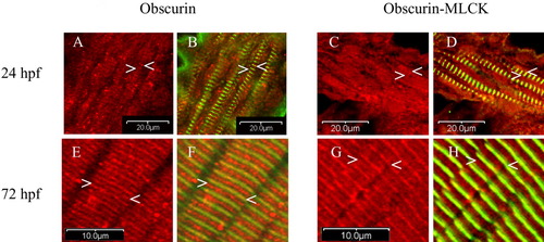
Cellular distribution of obscurin and obscurin-MLCK in zebrafish skeletal muscle during development. Embryos were fixed at 24 (A-D), and 72 (E-H) hpf and co-immunolabeled with antibodies to the ankyrin binding domain of obscurin (Ank) (red: A,B,E,F) or the carboxy terminal kinase domain of obscurin-MLCK (link7) (red: C,D,G,H) and α-actinin (green: B,D,F,H). At 24 hpf, while most of the cellular obscurin and obscurin-MLCK remains diffusely localized, some begins to organize around the Z (A-D:<) and M bands (A-D:>) of the maturing myofibrils. It is important to note that all myofibrils with a striated pattern of α-actinin staining also demonstrate organization of some of the obscurin and obscurin-MLCK around the M and Z bands. Later in development, by 72 hpf, both obscurin and obscurin-MLCK demonstrate a more distinct striated pattern with obscurin more concentrated at the M bands (E-H:>) and obscurin-MLCK at the Z bands (E-H:<). Similar results were obtained using antibodies to the amino terminal immunoglobulin domains of obscurin (4A8) and the internal kinase domain of obscurin-MLCK (SKII). Scale bars = 20 (A-D) and 10 (E-H) μm.
|
