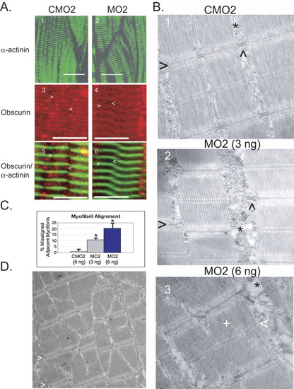
Obscurin depletion is associated with abnormalities of thick filament assembly and myofibril alignment in zebrafish embryos. At 72 hpf, embryos that had been injected with control (CMO2) or obscurin morpholino (MO2) were evaluated by immunohistochemical (A) and electron microscopic analysis (B). A: Control [CMO2 (6 ng); A.1,3,5] and obscurin morphant [MO2 (3 ng); A.2, (6 ng) A.4,6] embryos were hybridized with antibodies to α-actinin (A.1-2,5-6), and/or obscurin (Ank; A.3-6). A.5 and A.6 correspond to A.3 and A.4. Note that there irregularities of myofibril alignment and orientation in the skeletal muscle of the morphant embryos. The morphant embryos demonstrate a normal Z band structure and spacing as demonstrated by α-actinin localization, but there is decreased relative accumulation of obscurin at the M band (<) as opposed to the Z band (>) (compare A.4 and A.6 to A.3 and A.5). Scale bars = 20 μm. B: Electron micrographs of skeletal muscle from control [CMO2 (6 ng); B.1] and obscurin morphant [3 ng (B.2) and 6 ng (B.3) of MO2] embryos. Note the irregular spacing and misalignment of adjacent myofibrils in the morphant embryos. The Z bands (>) of adjacent myofibrils do not align and the sarcoplasmic reticulum (*) appears disorganized, often lacking the well-ordered triads (∧) evident in the control embryos. At the higher morpholino dose, there was occasional disorganization of the thick filaments (+) with irregularity of the M bands (<). C: Myofibril misalignment in control and morphant embryos. The percentage of myofibrils that were not aligned in register with the adjacent myofibril within the same myocyte was significantly greater in morphant than control embryos (*t-test; P < 0.01) and increased with increasing morpholino dose. D: Areas of myofibril disarray were clustered with some areas demonstrating high rates of misalignment. Note the staircase appearance of the Z bands (>) in the skeletal muscle of this 72-hpf embryo injected with 6 ng of MO2.
|
