
Fig. 4
- ID
- ZDB-FIG-060609-2
- Publication
- Connors et al., 2006 - Temporal and spatial action of Tolloid (Mini fin) and Chordin to pattern tail tissues
- Other Figures
- All Figure Page
- Back to All Figure Page
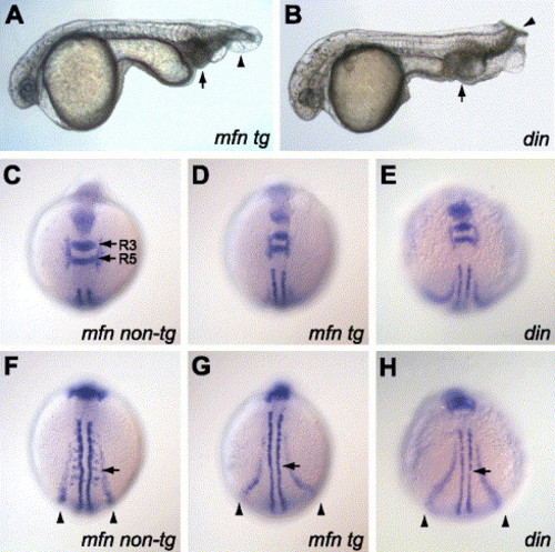
Overexpression of tolloid phenocopies the chordino mutant. A Tg(hsp70:tld) embryo heat-shocked during and after gastrulation to remove Chd activity (A) can phenocopy din/chd mutants (B). Eye and head size is reduced, the blood increased (arrow), and the tail fins duplicated (arrowhead). Dorsal-anterior views of a representative 7-somite stage non-transgenic mfn embryo (N = 37) (C), transgenic mfn embryo (N = 42) (D), or din homozygote (E) stained for myoD, krox20, gata1, and pax2.1. R3 and R5 refer to rhombomeres 3 and 5, respectively. (F–H) More posterior dorsal views of embryos shown in panels (C–E). Arrows indicate smaller somites, and arrowheads indicate more laterally located pronephric and blood precursor domains in transgenic and din mutants. |
| Genes: | |
|---|---|
| Fish: | |
| Condition: | |
| Anatomical Terms: | |
| Stage: | 5-9 somites |
Reprinted from Developmental Biology, 293(1), Connors, S.A., Tucker, J.A., and Mullins, M.C., Temporal and spatial action of Tolloid (Mini fin) and Chordin to pattern tail tissues, 191-202, Copyright (2006) with permission from Elsevier. Full text @ Dev. Biol.