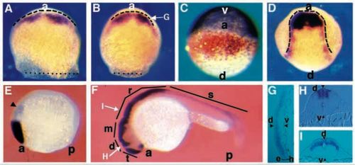
Zebrafish opl is expressed in the anterior neural plate and dorsal neural tube. Staged embryos were stained for opl RNA (purple, except in C, where it is orange) and gta3 RNA (purple in C) using whole-mount in situ hybridization. (A) Mid-gastrula (75% epiboly)embryo, dorsal view, dotted line indicates blastoderm margin, dashed line indicates anterior neural boundary. (B) Late gastrula (90% epiboly), dorsal view; dotted line indicates blastodermmargin, arrow indicates plane of section shown in G. (C) Late gastrula (90% epiboly), anterior view. (D) Early neurula (tailbud) embryo, dorsal view, dashed line outlines the neural plate. (E) Early somitogenesis (5 somites),side view, arrowhead indicates a weak posterior domain of staining. (F) Late somitogenesis (prim-5), side view, arrows indicate planes of section shown in H and I. (G) Transverse section through a late gastrula (90% epiboly) stage embryo as shown in D, showing that opl expression is confined to the epiblast (e) and is excluded from the hypoblast (h) (mesendoderm) layers (bracketed). Arrowheads mark dorsal and ventral limits of the blastoderm. (H,I) Transverse section through (H) the diencephalon, (I) the rhombencephalon; note restriction of expression to dorsal neural tube, arrowheads mark dorsal and ventral limits of the blastoderm. a, anterior; d, dorsal; v, ventral; p, posterior; t, telencephalon; d, diencephalon; m, mesencephalon; r, rhombencephalon; s, spinal cord.
|
