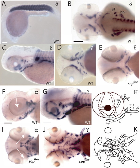
Expression of acetylcholine receptor (AChR) subunits in wild-type and sopfixe fish. A-E: Expression of the δ-subunit in wild-type (A-D) and sopfixe mutant (E) embryos. The δ-subunit is expressed exclusively in muscles. No differences in expression levels between wild-type and sopfixe mutants were detected (compare D with E; A, 24 hpf; B-E, 50 hpf). F,G,I,J: Comparison of the expression of the α1-subunit (F,I) and the γ-subunit (G,J) at 72 hpf. In contrast to all other subunits analyzed, the α1-subunit does not continue to be expressed in eye muscles after 50 hpf (compare F, I, arrow, with G,J). H,K: Schematic drawing of the muscles in the head in lateral (H) and ventral (K) views. ah, adductor hyoideus; am, adductor mandibulae; ao, adductor operculi; do, dilator operculi; hh, hyohyoideus; hp, hypaxial muscles; ih, interhyoideus; ima, intramandibular anterior; imp, intramandibular posterior; io, inferior oblique; ir, inferior rectus; lap, levator arcus palatini; lr, lateral rectus; mr, medial rectus; pf, pectoral fin; sh, sternohyoideus; so, superior oblique; tv, transversus ventralis. A,C,F,G,H: Lateral view; B: dorsal view; D,E,I,J,K: ventral view. Scale bars = 80 μm in A, 60 μm in B-E, 50 μm in F-J.
|
