
Fig. 5
- ID
- ZDB-FIG-051215-4
- Publication
- Etard et al., 2005 - Mutation in the delta-subunit of the nAChR suppresses the muscle defects caused by lack of Dystrophin
- Other Figures
- All Figure Page
- Back to All Figure Page
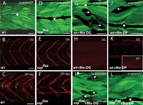
sopfixe suppresses the muscle defects in embryos lacking Dystrophin. A-C: Uninjected wild-type embryos showing expression of the α-actin:GFP transgene (A) or expression of dystroglycan (DG, B) or the combination of Dystrophin (DP) immunofluorescence and α-bungarotoxin staining (C). D-F: sopfixe mutants showing expression of the α-actin:GFP transgene (D) or expression of DG (E) or the combination of DP immunofluorescence and α-bungarotoxin staining (F). Lack of α-bungarotoxin in sopfixe does not affect the pattern of DG or DP expression (compare B,C with E,F). G,H: Wild-type embryos injected with the morpholino against DG (Mo-DG). G: Removal of DG causes detachment of the α-actin:gfp labeled myofibrils from the vertical myosepta (arrows). H: The injected Mo-DG completely abolishes DG expression. I: sopfixe embryo injected with Mo-DG shows a similar pattern of muscle defects as wild-type embryos, in which DG was knocked down, indicating that sopfixe does not suppress the muscle phenotype in Mo-DG morphants. J,K: Embryos in which DP was knocked down show detachment of myofibrils (J) and also a reduction in DG as well as DP staining at the vertical myosepta (K, and insert in K, respectively). L: sopfixe mutant embryos in which DP was knocked down do not show detachment of myofibrils from the vertical myoseptum. Thus, sopfixe suppresses the muscle defects in Mo-DP morphants. A,B, D,E,G-L: 72 hours postfertilization (hpf); C,F: 24 hpf embryos. Asterisk, horizontal myosepta. GFP, green fluorescent protein. Scale bars = 30 μm in A,B,D,E,G-L; 25 μm in C,F. |