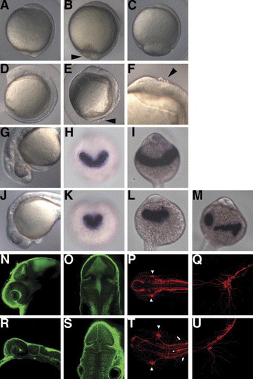
The rk3 mutant shows cell detachment, epiboly delay, and abnormalities in the formation of neural tissues and the hatching gland. (A–C) Morphology of the wild-type (A) and rk3 homozygous mutant embryos (B, C) at 10 hpf (bud stage for wild-type embryos). Lateral views with dorsal to the right. A group of the embryos completed epiboly at 10 hpf, but showed an irregular embryonic surface, distorted dorsal midline, and dissociation of cells (B, arrowhead). Severely affected embryos showed a delay in epiboly (C). The severity of the mutant phenotypes varied depending on the mating pairs and culture temperature (Fig. 2). (D–F) Morphology of the wild-type (D) and rk3 mutant embryos (E, F) at the segmentation stage (10-somite stage). (F) High magnification of the anterior region of the mutant embryo. The mutant embryos had a shorter anterior–posterior axis and wider somites than wild-type embryos, and cells that were dissociated from the embryos (indicated by arrowheads in E and F). The embryos often underwent yolk lysis and died during the segmentation period. (G,J) Morphology of the wild-type (G) and rk3 mutant embryos (J) at the pharyngula stage (24 hpf). Anterior is to the left. (I, L). In the mutant embryos, the anterior neural tissue was flattened and the hatching gland stayed in the midline and did not expand laterally. (H, I, K–M) Expression of the hatching gland marker hgg1 in the wild-type (H, I) and the rk3 mutant embryos (K–M) at the early segmentation stage (H, K) and at 24 hpf (I, L, M). Anterior views. The polster was wide in the medio-lateral axis in the wild-type embryos (H), but stayed in the midline at the segmentation stage in the rk3 mutant embryos (K). The hatching gland stayed on the front of the head (L), or was broken apart and mislocated (M) in the rk3 mutant embryos. (N, O, R, S) Bodipy-ceramide staining of the wild-type (N, O) and the rk3 mutant embryos (R, S) at 30 hpf. Confocal microscopic sagittal (N, R) and cross (O, S) sections. The rk3 mutant embryos showed a flattened nervous structure. (R, S, V, W) Immunostaining with anti-acetylated tublin at 30 hpf in wild-type (P, Q) and rk3 mutant (T, U) embryos. Dorsal views with anterior to the left (P, T). High-magnification views of the trigeminal ganglia and lateral views with anterior to the left (Q, U). The trigeminal ganglia (arrowheads in P, T) were located more laterally, and the neurons in the trigeminal ganglia were scattered or loosely organized, and their axons were disorganized in the rk3 mutant embryos. Abnormal positioning of Rohon-Beard neurons (arrows) and aberrant axon tracts in the dorsal midline (dot in T) were observed in the posterior hindbrain and the anterior spinal cord in the rk3 mutant embryos.
|
