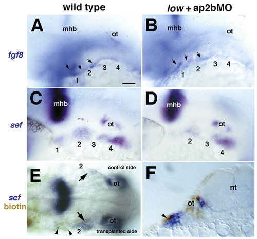
tfap2a and tfap2b are required for Fgf signaling in NCCs. Lateral views, anterior towards the left. (A,B) At 32 hpf, fgf8 mRNA is detected in wild type in the ectoderm covering the arches, in addition to the mid-hindbrain boundary (mhb); this expression remains in AP2a/b-deficient embryos. Arrows in A,B indicate ectodermal expression of fgf8. (C,D) At 28 hpf in wild type, sef mRNA is detected in NCCs; this expression is severely reduced in AP2a/b-deficient embryos. (E,F) Mosaic embryos in which biotin-labeled, wild-type ectodermal cells (brown, arrows) were transplanted on one side of the head and partially restore sef expression (blue). (E) Dorsal view, anterior towards the left showing transplanted side with substantially increased levels of sef expression, and lack of expression on the contralateral, control side (arrows). (F) A transverse section through the embryo shown in E at the level of the hyoid arch showing grafted wild-type ectoderm (brown) and rescued sef expression in underlying NC (blue). Arrowhead in F indicates transplanted ectoderm. m, mandibular arch; h, hyoid arch; b, branchial arches. Scale bars: 50 µm.
|
