
Fig. 4
- ID
- ZDB-FIG-050628-2
- Publication
- Marza et al., 2005 - Developmental expression and nutritional regulation of a zebrafish gene homologous to mammalian microsomal triglyceride transfer protein large subunit
- Other Figures
- All Figure Page
- Back to All Figure Page
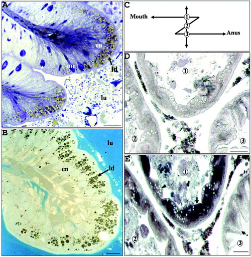
Intestinal mucosa of fed adult zebrafish. A,B: Histological semithin sections of the anterior intestine were stained with toluidine blue (A) or with the neutral lipid-specific stain Sudan black B (B). In both cases, the intestinal mucosa showed enterocytes (en) containing large lipid droplets (ld). C: Schema of adult zebrafish intestine. From the mouth to the anus, two intestinal loops are observed and transverse sections at loop levels give three different intestinal rings, numbered 1 to 3. D,E: The localization of mtp transcripts was performed in the intestine by in situ hybridizations with digoxigenin-labeled sense (D) and antisense (E) riboprobes on parallel transverse intestinal sections of an 8-month fed young adult female at the level indicated by a vertical line in C. Rings 1 and 2 are representative of the proximal and distal parts of the anterior intestine, respectively. Ring 3 could be attributed to the posterior part of the intestine due to the presence of a large amount of vacuoles in the supranuclear hyaloplasm (arrow with broken line) of enterocytes. No staining signal was observed by using the sense probe. A strong mtp hybridization signal was observed at ring 1 and, to a lesser extent, at rings 2 and 3. The staining signal was restricted to the enterocytes (arrow), whereas the mucous cells (arrowhead) were not labeled. lu, lumen; mc, mucous cells. Scale bars = 30 μm in A, 15 μm in B, 100 μm in D,E. |