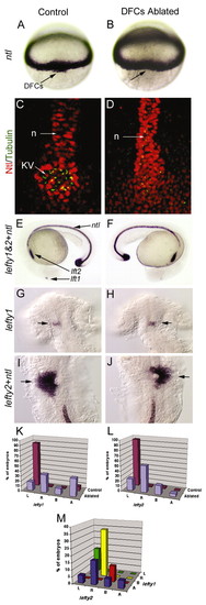
Laser ablation of DFCs alters LR patterning without affecting development of the midline. Unablated control embryos expressed ntl in DFCs (arrow) at 60% epiboly (A) and in the notochord at 24 hours post-fertilization (E). Laser ablation eliminated DFCs (arrow, dorsal view at 60% epiboly) but did not alter ntl expression in equatorial mesoderm (B) or in notochord and tailbud at 24 hpf (F). Control embryos (C) and DFC-ablated embryos (D) were immunostained with anti-acetylated Tubulin antibodies (green) to detect cilia and anti-ntl antibodies (red) to detect the notochord (n) and Kupffers vesicle (KV). Both control and DFC-ablated embryos showed contiguous ntl staining in the notochord, even in embryos (n=2/6) that failed to form KV (D). (G-J) In control embryos, we observed normal expression of lft1 (arrow) in the left dorsal diencephelon (G) and lft2 (arrow) in the left heart primordia (I). In DFC-ablated embryos, lft1 (arrow) in the diencephalon (H) and lft2 (arrow) in the heart primordia (J) were frequently reversed. (K-M) Analysis of lft1 expression in the diencephalon (K) and lft2 expression in the heart primordia (L) in control (n=71) and DFC-ablated (n=17) embryos. (M) Analysis of lft1 expression plotted against lft2 expression in DFC-ablated embryos indicated that the predominant class of DFC-ablated embryos displayed reversal of both brain and heart markers (yellow bar). L, left; R, right; B, bilateral; A, absent gene expression.
|
