
- Title
-
Altered GABAergic, glutamatergic and endocannabinoid signaling is accompanied by neuroinflammatory response in a zebrafish model of social withdrawal behavior
- Authors
- Perdikaris, P., Dermon, C.R.
- Source
- Full text @ Front. Mol. Neurosci.
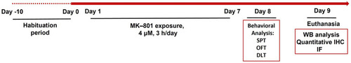
Schematic representation of the experimental timeline including the methodological approaches used to evaluate zebrafish social and anxiety-like behavior following sub-chronic MK-801 treatment. SPT, social preference test; OFT, open field test; DLT, dark–light test; WB, western blot; IHC, immunohistochemistry; IF, immunofluorescence. |
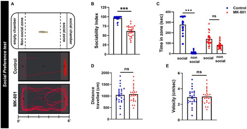
MK-801-treated zebrafish display social impairments. (A) Diagram of social preference test and representative behavioral trajectories of control and MK-801-treated zebrafish. (B–E) Social and locomotor parameters of control and MK-801-treated fish as estimated in the social preference test. (B) Sociability index, (C) time spent in social (S) and non-social (NS) zone, (D) distance traveled, and |
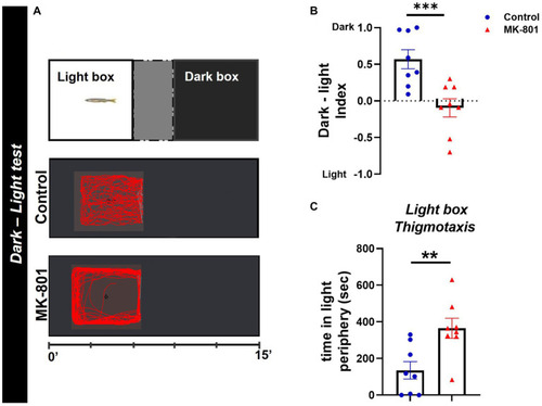
MK-801-treated zebrafish exhibit abnormal dark avoidance behavior. (A) Diagram of dark–light test and representative behavioral trajectories of control and MK-801-treated zebrafish. (B,C) Anxiety parameters of Control and MK-801-treated zebrafish as estimated in the dark–light test. (B) Dark–light index and (C) light box thigmotaxis. n = 8 per experimental group. Data are expressed as mean ± SEM. **p ≤ 0.01, ***p ≤ 0.001, compared to control group. |
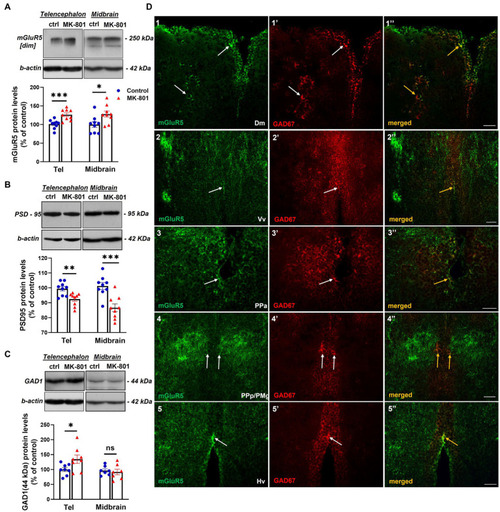
MK-801-treated zebrafish are characterized by altered expression levels of proteins involved in glutamatergic and GABAergic neurotransmission (A–C) Representative blots and quantification of the relative expression levels of (A) mGluR5, (B) PSD-95 and (C) GAD1. n = 6–7 (GAD1) or n = 9 (mGlur5, PSD-95) per experimental group. Data are expressed as mean ± SEM. *p ≤ 0.05, **p ≤ 0.01, ***p ≤ 0.001, ns, non-significant, compared to control group. (D) Immunofluorescent microphotographs of selected transverse sections showing colocalization of mGluR5 with GAD1 immunoreactive cells within different areas of the SDMN. (D1–1’’) medial zone of the dorsal telencephalic area (Dm), (D2–2’’)- ventral nucleus of the ventral telencephalic area (Vv), (D3–3’’) parvocellular preoptic nucleus, anterior part (PPa), (D4–4’’) parvocellular preoptic nucleus, posterior part/gigantocellular part of magnocellular preoptic nucleus (PPp/PMg), (D5–5’’) ventral zone of periventricular hypothalamus (Hv). Arrows indicate examples of colocalization. Microphotographic images are representative of both control (D1–1”,D3–3”,D4–4”,D5–5”) and MK-801-treated (D2–2’’) fish. Scale bar: 25 μm. |
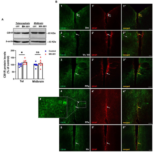
Altered endocannabinoid signaling in telencephalon of MK-801-treated zebrafish. (A) Representative blots and quantification of the relative expression levels of CB1R in telencephalon and midbrain. n = 8 per experimental group. Data are expressed as mean ± SEM. *p ≤ 0.05, ns, non-significant, compared to control group. (B1–5’’) Immunofluorescent microphotographs of selected transverse sections showing colocalization of CB1R with GFAP immunoreactive cells in the periventricular zone within different nodes of the SDMN. (B1–1’’) medial zone of the dorsal telencephalic area (Dm), (B2–2’’) ventral nucleus of the ventral telencephalic area/dorsal nucleus of the ventral telencephalic area (Vv/Vd), (B3–3’’) parvocellular preoptic nucleus, anterior part (PPa), (B4–4’’) parvocellular preoptic nucleus, posterior part (PPp), (B5–5’’) ventral zone of periventricular hypothalamus (Hv). Arrows indicate examples of colocalization, dashed arrows point to single positive cells. Within brain parenchyma, CB1R-immunostaining is intensively present in punctate structures around or in close proximity with cell somata (B4#). Microphotographic images are representative of MK-801-treated fish. Scale bar: 25 μm. |
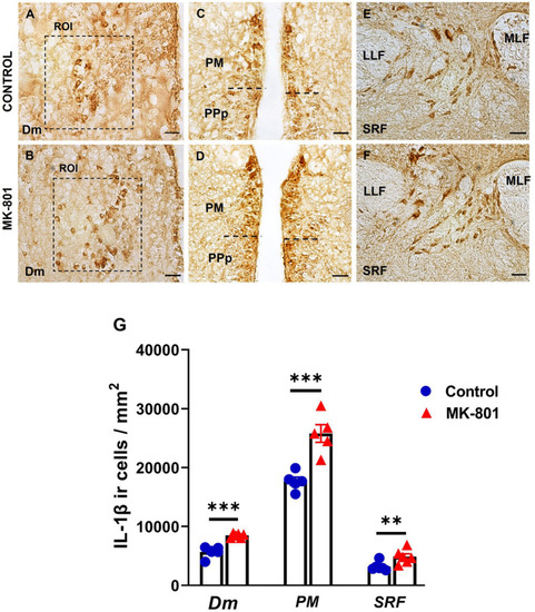
Excessive IL-1β expression within the SDMN and SRF, in MK-801 treated zebrafish displaying social withdrawal and anxiety-like behavior. (A–F) Representative staining for IL-1β in (A,B) medial zone of the dorsal telencephalic area (Dm), (C,D) magnocellular preoptic nucleus and (E,F) superior reticular formation (SRF) of control and MK-801 treated fish. Scale bar: 25 μm. (G) The density of IL-1β positive cells were significantly increased in MK-801 treated zebrafish, in Dm, PM and SRF, compared to control group. Bars represent the number (mean ± SEM) of IL-1β positive cells. n = 5 per experimental group. **p ≤ 0.01, ***p ≤ 0.001, compared to control group. |
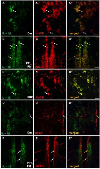
Expression of IL-1β in neurons and/or astrocytes within areas of the SDMN, in MK-801-treated zebrafish. (A–E’’) Immunofluorescent microphotographs of selected transverse sections showing colocalization of IL-1β with HuC/D (A–C”), or GFAP (D–E”) in medial zone of the dorsal telencephalic area (Dm) (A–A”,D–D”), parvocellular preoptic nucleus, posterior part/magnocellular preoptic nucleus (PPp, PM) (B–B”,E–E”) and superior reticular formation (SRF) (C). Arrows indicate examples of colocalization, dashed arrows point to single positive cells. Microphotographic images are representative of MK-801-treated fish, n = 5. Scale bar: 25 μm. ROI, region of interest. |
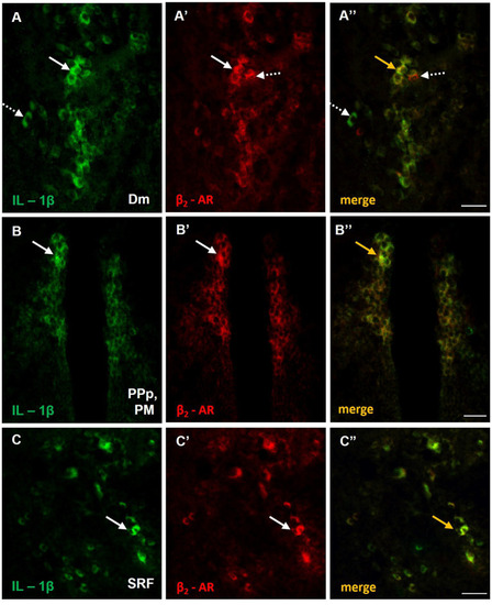
Possible noradrenergic regulation of IL-1β expression through β2-adrenoceptors (β2-ARs) activation within areas of the SDMN. (A–C’’) Immunofluorescent microphotographs of selected transverse sections showing colocalization of IL-1β with β2-ARs in (A–A’’) medial zone of the dorsal telencephalic area (Dm), (B–B’’) parvocellular preoptic nucleus, posterior part/magnocellular preoptic nucleus (PPp, PM), and (C–C’’) superior reticular formation (SRF). Arrows indicate examples of colocalization, dashed arrows point to single positive cells. Microphotographic images are representative of MK-801-treated fish, n = 5. Scale bar: 25 μm. |
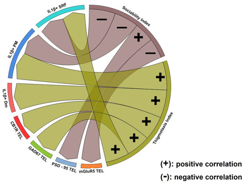
Chord diagram obtained from Spearman’s correlation analysis showing the relationship between social withdrawal and anxiety-like behavior, excitation/inhibition imbalance and excessive IL-1β expression. Links indicate strong positive (+) and negative (−) significant correlations, with “r” values above 0.6 or below −0.6, respectively. Notice that glutamatergic dysfunction was associated with social withdrawal behavior whereas GABAergic and endocannabinoid system dysfunction was associated with increased anxiety. Excessive IL-1β expression in Dm was associated with anxiety-like behavior while in PM and SRF was linked both to social withdrawal and increased anxiety. p ≤ 0.05. |