
- Title
-
The Spike protein of SARS-CoV-2 signals via Tlr2 in zebrafish
- Authors
- Tyrkalska, S.D., Martínez-López, A., Pedoto, A., Candel, S., Cayuela, M.L., Mulero, V.
- Source
- Full text @ Dev. Comp. Immunol.
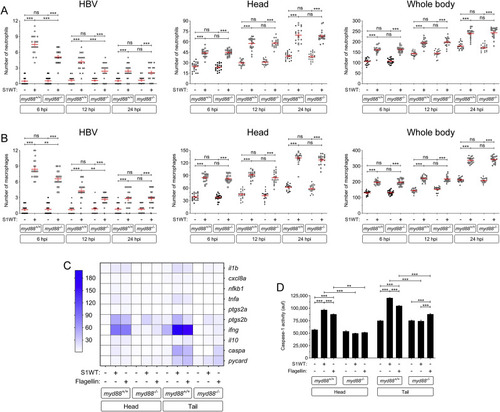
Myd88 is required for hyperinflammation but dispensable for emergency myelopoiesis induced by S1WT. Recombinant S1WT (+) or vehicle (−) were injected in the hindbrain ventricle (HBV) of 2 dpf Myd88-deficient Tg(mpx:eGFP) (A) or wild type (B–D) larvae. Neutrophil (A) and macrophage (neutral red positive cells) (B) recruitment and number were analyzed at 6, 12 and 24 hpi by fluorescence (A) or brightfield (B) microscopy, the transcript levels of the indicated genes were analyzed at 12 hpi by RT-qPCR in larval head and tail (C), and caspase-1 activity was determined at 24 hpi using a fluorogenic substrate (D). Each dot represents one individual and the mean ± S.E.M. for each group is also shown. P values were calculated using one-way ANOVA and Tukey multiple range test. RT-qPCR data are depicted as a heat map in C with higher expression shown in darker color. ns, not significant, **p ≤ 0.01, ***p ≤ 0.001. auf, arbitrary units of fluorescence. |
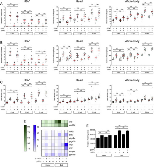
Il1b signaling is not involved in S1WT-induced hyperinflammation in zebrafish. One-cell stage zebrafish eggs of Tg(lyz:dsRED2) (A), Tg(mfap4:mCherry) (B), Tg(NFkB-RE:eGFP) (C) and wild type (D, E) were microinjected with control or il1b crRNA/Cas9 complexes. At 2 dpf, recombinant S1WT (+) or vehicle (−) were injected in the hindbrain ventricle (HBV) of control and Il1b-deficient larvae. Neutrophil (A) and macrophage (B) recruitment and number, and Nfkb activation (C) were analyzed at 6, 12 and 24 hpi by fluorescence microscopy, the transcript levels of the indicated genes were analyzed at 12 hpi by RT-qPCR (D), and caspase-1 activity was determined at 24 hpi using a fluorogenic substrate (E). Each dot represents one individual and the mean ± S.E.M. for each group is also shown. RT-qPCR data are depicted as a heat map in D with higher expression shown in darker color. P values were calculated using one-way ANOVA and Tukey multiple range test. ns, not significant, *≤p0.05, **p ≤ 0.01, ***p ≤ 0.001. auf, arbitrary units of fluorescence. |

Tlr2 mediates the S1WT-induced hyperinflammation in zebrafish. One-cell stage zebrafish eggs of Tg(lyz:dsRED2) (A), Tg(mfap4:mCherry) (B), Tg(NFkB-RE:eGFP) (C) and wild type (D, E) were microinjected with control or tlr2 crRNA/Cas9 complexes. At 2 dpf, recombinant S1WT (+) or vehicle (−) were injected in the hindbrain ventricle (HBV) of control and Tlr2-deficient larvae. Neutrophil (A) and macrophage (B) recruitment and number, and Nfkb activation (C) were analyzed at 6, 12 and 24 hpi by fluorescence microscopy, the transcript levels of the indicated genes were analyzed at 12 hpi by RT-qPCR (D), and caspase-1 activity was determined at 24 hpi using a fluorogenic substrate (E). Each dot represents one individual and the mean ± S.E.M. for each group is also shown. RT-qPCR data are depicted as a heat map in D with higher expression shown in darker color. P values were calculated using one-way ANOVA and Tukey multiple range test. ns, not significant, *≤p0.05, **p ≤ 0.01, ***p ≤ 0.001. auf, arbitrary units of fluorescence. |