
- Title
-
Plexind1 negatively regulates zebrafish lymphatic development
- Authors
- Britto, D.D., He, J., Misa, J.P., Chen, W., Kakadia, P.M., Grimm, L., Herbert, C.D., Crosier, K.E., Crosier, P.S., Bohlander, S.K., Hogan, B.M., Hall, C.J., Torres-Vázquez, J., Astin, J.W.
- Source
- Full text @ Development

plxnd1 mutants have misaligned ISLVs and aberrant facial lymphatic branches. (A-D′) Confocal images of the trunk (A-A″,B-B″) and facial (C,D) lymphatics in lyve1b:DsRed (white); kdrl:EGFP (green) larvae at 6 dpf. Yellow arrowheads indicate ISLVs that are not aligned with an ISV (B,B″) or aberrant facial lymphatic branches (D) in plxnd1nz75 larvae. For clarity, EGFP-positive vessels in the head are not displayed. Schematics highlight the misaligned ISLVs (A‴,B‴) or aberrant facial lymphatics (C′,D′) in each corresponding confocal image. (E,F) Quantitation of the number of misaligned ISLVs (E) and aberrant facial lymphatic branches (F). P>0.05 (n.s., not significant), *P<0.05, **P<0.01 (Brown-Forsythe, and Welch ANOVA and Kruskal–Wallis test); data are mean±s.d. Scale bars: 50 µm. ISLV, intersegmental lymphatic vessel. Numbers in graphs represent numbers of larvae. |

plxnd1 is required for the alignment of developing ISLVs to blood vessels. (A-H) Still images from time-lapse movies of ISLV growth between 60 and 70 hpf in lyve1b:DsRed; kdrl:EGFP wild type (A-D) and plxnd1nz75 (E-H) larvae showing blood vessels (green) lyve1b-expressing vessels (white) from Movies 1 and 2, respectively. Yellow arrowheads indicate the distal tip of each ISLV. (I,J) Traces of ISLV growth paths for wild type (I) and plxnd1 (J) (n=4). ISLVs were traced dorsally from when they left the horizontal myoseptum. (K) Quantitation of the percentage of time the ISLVs are aligned with a blood vessel (n=4). (L) Quantitation of the number of ISLV branching events (n=4). (M) Quantitation of the ISLV meandering index (n=4). *P<0.05 (Mann–Whitney test); data are mean±s.d. Scale bar: 50 µm. ISLV, intersegmental lymphatic vessel. |
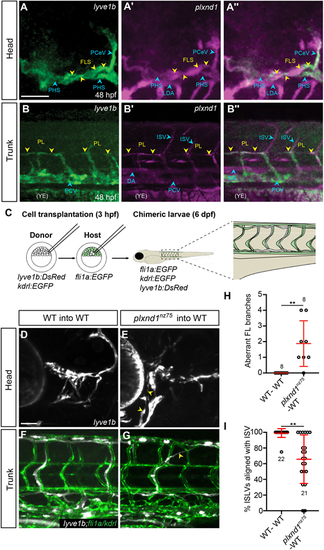
plxnd1 acts cell-autonomously to guide lymphatic vessel growth. (A-B″) Confocal images of the lyve1b:EGFP expression (anti-EGFP, green) (A,B), plxnd1 expression (anti-plxnd1, magenta) (A′,B′) or both (A″,B″) in the head (A,A″) or trunk (B,B″) of 48 hpf lyve1b:EGFP larvae. Yellow arrowheads indicate either the facial lymphatic sprout (A) or the parachordal LECs (B). Blue arrowheads indicate blood vessels. There is non-specific staining in the yolk extension (YE). (C) Schematic of the transplantation protocol. Cells were transplanted from 3 hpf lyve1b:DsRed; kdrl:EGFP donor embryos to 3 hpf fli1a:EGFP host embryos, resulting in chimeric larva with host-derived blood and lymphatic vessels indicated in light green and donor-derived lymphatic vessels indicated in white. (D,E) Confocal images of the facial vasculature in chimeric animals at 6 dpf showing donor-derived LECs (white) from either wild-type (D) or plxnd1nz75 (E) donors. For clarity, EGFP-positive vessels are not displayed. Aberrant plxnd1nz75 facial lymphatic vessels are highlighted by yellow arrowheads in E. (F,G) Confocal images of the trunk vasculature in chimeric animals at 6 dpf showing donor-derived LECs (white) from either wild-type (F) or plxnd1nz75 (G) donors. Note the misaligned plxnd1nz75 vessel highlighted by the yellow arrowhead in G. (H,I) Quantitation of aberrant facial lymphatics (H) or the number of misaligned ISLVs (I) at 6 dpf. **P<0.01 (Mann–Whitney test); data are mean±s.d. Scale bars: 50 µm. DA, dorsal aorta; FLS, facial lymphatic sprout; ISV, intersegmental vessel; ISLV, intersegmental lymphatic vessel; LDA, lateral dorsal aorta; PCV, posterior cardinal vein; PCeV, posterior cerebral vein; PL, parachordal LEC; PHS, primary head sinus; YE, yolk extension. Numbers in graphs represent numbers of larvae. |

plxnd1 mutants display facial lymphatic hyperplasia. (A,B) Confocal images of the facial vasculature showing the lyve1b-expressing vessels (white) in 6 dpf lyve1b:DsRed; fli1a:nlsEGFP wild-type (A) or plxnd1nz75 (B) larvae. (A′,B′) Areas outlined in A,B showing LECs within the OLV (lyve1b, magenta; fli1a:nlsEGFP, green). Yellow arrowheads indicate OLV nuclei. (C) Quantitation of facial LEC number at 6 dpf. (D) Quantitation facial LEC number from 3 to 6 dpf. (E) Quantitation of trunk LEC number at 6 dpf. P>0.05 (not significant), *P<0.05, **P<0.01 (one-way ANOVA); data are mean±s.d. Scale bar: 50 µm. LEC, lymphatic endothelial cell; OLV, otolithic lymphatic vessel. Numbers in graphs represent numbers of larvae. |
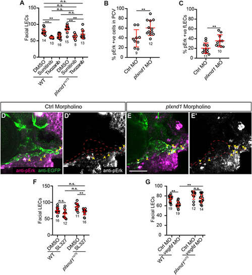
plxnd1 mutants have elevated Vegfr/Erk signalling in the facial lymphatics. (A) Quantitation of facial LEC number in 6 dpf larvae treated from 3 to 6 dpf with either DMSO, 1 µM sunitinib or 2 nM tivozanib. (B,C) Quantitation of the proportion of pErk-positive cells within the PCV at 36 hpf (B) or the facial LECs at 3 dpf (C). (D-E′) Confocal images of anti-pErk (magenta) and anti-EGFP (green) staining in the head at 3 dpf of either control (D,D′) or plxnd1 (E,E′) lyve1b:EGFP morphant larvae. (D′,E′) Anti-pErk staining only. Yellow arrowheads indicate the pErk cells within the facial lymphatics. (F) Quantitation of facial LEC number in 6 dpf larvae treated from 3 to 6 dpf with either DMSO or 5 µM SL327. (G) Quantitation of facial LEC number in lyve1b:DsRed; fli1a:nlsEGFP larvae injected with either control or vegfd morpholinos. P>0.05 (not significant), *P<0.05, **P<0.01 (unpaired t-tests, Kruskal–Wallis and ANOVA); data are mean±s.d. Scale bar: 50 µm. LEC, lymphatic endothelial cell. Numbers in graphs represent numbers of larvae. |
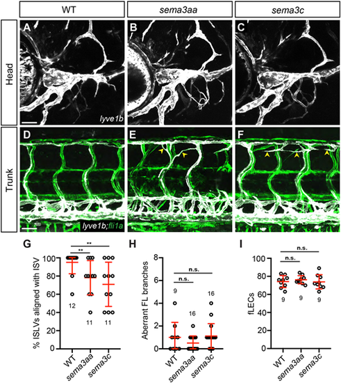
Semaphorin 3 ligands regulate trunk lymphatic guidance. (A-F) Confocal images of the head (A-C) or trunk (D-F) vasculature showing the blood (green) and lyve1b-expressing vessels (white) in 6 dpf lyve1b:DsRed; fli1a:EGFP wild type (A,D) and sema3aasa10241 (B,E) or sema3csa15161 (C,F) mutants. Yellow arrowheads indicate misaligned ISLVs. (G) Quantitation of misaligned ISLVs at 6 dpf. (H) Quantitation of aberrant facial lymphatics at 6 dpf. (I) Quantitation of facial LEC number at 6 dpf. P>0.05 (not significant), **P<0.01 (by Mann–Whitney test); data are mean±s.d. Scale bar: 50 µm. ISLV, intersegmental lymphatic vessel. Numbers in graphs represent numbers of larvae. |

Plxnd1 signalling regulates lymphatic vessel guidance and growth. Model of Plxnd1-mediatated regulation of lymphatic development. (A) In the head, Plxnd1 signalling antagonises Vegfr/Erk signalling to prevent facial lymphatic hyperplasia. It also prevents aberrant cell migration. The role and identity of semaphorin ligands in facial lymphatic development are yet to be determined. (B) In the trunk, Plxnd1 signalling within developing ISLVs, likely triggered by Sema3aa/Sema3c ligands, prevents migration into the somites. |