
|
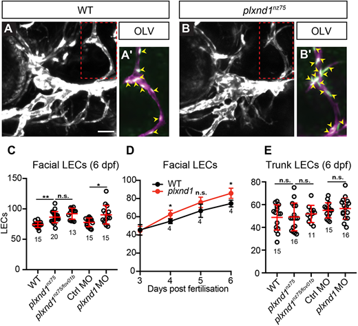
Fig. 4 plxnd1 mutants display facial lymphatic hyperplasia. (A,B) Confocal images of the facial vasculature showing the lyve1b-expressing vessels (white) in 6 dpf lyve1b:DsRed; fli1a:nlsEGFP wild-type (A) or plxnd1nz75 (B) larvae. (A′,B′) Areas outlined in A,B showing LECs within the OLV (lyve1b, magenta; fli1a:nlsEGFP, green). Yellow arrowheads indicate OLV nuclei. (C) Quantitation of facial LEC number at 6 dpf. (D) Quantitation facial LEC number from 3 to 6 dpf. (E) Quantitation of trunk LEC number at 6 dpf. P>0.05 (not significant), *P<0.05, **P<0.01 (one-way ANOVA); data are mean±s.d. Scale bar: 50 µm. LEC, lymphatic endothelial cell; OLV, otolithic lymphatic vessel. Numbers in graphs represent numbers of larvae.